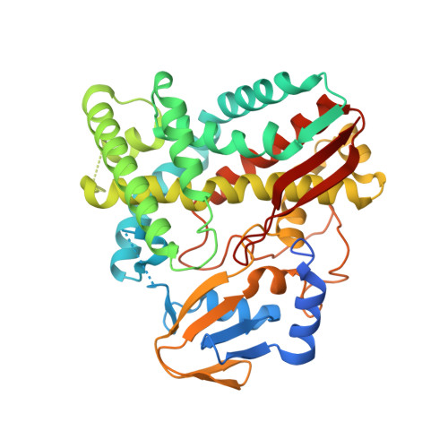
4RM4

The crystal structure of the versatile cytochrome P450 enzyme CYP109B1 from Bacillus subtilis


Experimental Data Snapshot

  • Method: X-RAY DIFFRACTION
  • Resolution: 1.77 Å
  • R-Value Free: 0.253 
  • R-Value Work: 0.202 
  • R-Value Observed: 0.205 

Starting Model: experimental
View more details

wwPDB Validation   3D Report Full Report


Ligand Structure Quality Assessment 


This is version 1.2 of the entry. See complete history


Literature

The crystal structure of the versatile cytochrome P450 enzyme CYP109B1 from Bacillus subtilis.

Zhang, A.Zhang, T.Hall, E.A.Hutchinson, S.Cryle, M.J.Wong, L.L.Zhou, W.Bell, S.G.

(2015) Mol Biosyst 11: 869-881

  • DOI: https://doi.org/10.1039/c4mb00665h
  • Primary Citation of Related Structures:  
    4RM4

  • PubMed Abstract: 

    The crystal structure of the versatile CYP109B1 enzyme from Bacillus subtilis has been solved at 1.8 Å resolution. This is the first structure of an enzyme from this CYP family, whose members are prevalent across diverse species of bacteria. In the crystal structure the enzyme has an open conformation with an access channel leading from the heme to the surface. The substrate-free structure reveals the location of the key residues in the active site that are responsible for binding the substrate in the correct orientation for regioselective oxidation. Importantly, there are significant differences among these residues in members of the CYP109 and closely related CYP106 families and these likely account for the variations in substrate binding and oxidation profiles observed with these enzymes. A whole-cell oxidation biosystem was developed, which contains CYP109B1 and a phthalate family oxygenase reductase (PFOR), from Pseudomonas putida KT24440, as the electron transfer partner. This electron transfer system is able to support CYP109B1 activity resulting in the regioselective hydroxylation of both α- and β-ionone in vivo and in vitro. The PFOR is therefore a versatile electron transfer partner that is able to support the activity of CYP enzymes from other bacterium. The crystal structure of CYP109B1 has a positively charged proximal face and this explains why it can interact with PFOR and adrenodoxin which are predominantly negatively charged around their [2Fe-2S] clusters.


  • Organizational Affiliation

    College of Life Sciences, Nankai University, Tianjin 300071, China. zhouwh@nankai.edu.cn.


Macromolecules
Find similar proteins by:  (by identity cutoff)  |  3D Structure
Entity ID: 1
MoleculeChains Sequence LengthOrganismDetailsImage
Cytochrome P450396Bacillus subtilis subsp. subtilisMutation(s): 0 
Gene Names: BSU12210genomic DNAyjiB
EC: 1.14
UniProt
Find proteins for O34374 (Bacillus subtilis (strain 168))
Explore O34374 
Go to UniProtKB:  O34374
Entity Groups  
Sequence Clusters30% Identity50% Identity70% Identity90% Identity95% Identity100% Identity
UniProt GroupO34374
Sequence Annotations
Expand
  • Reference Sequence
Small Molecules
Ligands 1 Unique
IDChains Name / Formula / InChI Key2D Diagram3D Interactions
HEM
Query on HEM

Download Ideal Coordinates CCD File 
B [auth A]PROTOPORPHYRIN IX CONTAINING FE
C34 H32 Fe N4 O4
KABFMIBPWCXCRK-RGGAHWMASA-L
Experimental Data & Validation

Experimental Data

  • Method: X-RAY DIFFRACTION
  • Resolution: 1.77 Å
  • R-Value Free: 0.253 
  • R-Value Work: 0.202 
  • R-Value Observed: 0.205 
  • Space Group: P 1 21 1
Unit Cell:
Length ( Å )Angle ( ˚ )
a = 53.996α = 90
b = 67.573β = 113.04
c = 56.406γ = 90
Software Package:
Software NamePurpose
HKL-2000data collection
PHASESphasing
PHENIXrefinement
HKL-2000data reduction
HKL-2000data scaling

Structure Validation

View Full Validation Report


Ligand Structure Quality Assessment 


Entry History 

Deposition Data

Revision History  (Full details and data files)

  • Version 1.0: 2015-02-04
    Type: Initial release
  • Version 1.1: 2015-03-04
    Changes: Database references
  • Version 1.2: 2023-09-20
    Changes: Data collection, Database references, Derived calculations, Refinement description