


Crystal structure of human dihydroorotate dehydrogenase (DHODH) with DH03A048
Zhu, L., Ren, X., Zhu, J., Li, H.To be published.
Experimental Data Snapshot
Starting Model: experimental
View more details
Entity ID: 1 | |||||
|---|---|---|---|---|---|
| Molecule | Chains | Sequence Length | Organism | Details | Image |
| Dihydroorotate dehydrogenase (quinone), mitochondrial | 390 | Homo sapiens | Mutation(s): 0 Gene Names: DHODH EC: 1.3.5.2 | 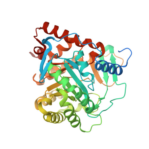 | |
UniProt & NIH Common Fund Data Resources | |||||
PHAROS: Q02127 GTEx: ENSG00000102967 | |||||
Entity Groups | |||||
| Sequence Clusters | 30% Identity50% Identity70% Identity90% Identity95% Identity100% Identity | ||||
| UniProt Group | Q02127 | ||||
Sequence AnnotationsExpand | |||||
Reference Sequence | |||||
| Ligands 3 Unique | |||||
|---|---|---|---|---|---|
| ID | Chains | Name / Formula / InChI Key | 2D Diagram | 3D Interactions | |
| FMN Download:Ideal Coordinates CCD File | B [auth A] | FLAVIN MONONUCLEOTIDE C17 H21 N4 O9 P FVTCRASFADXXNN-SCRDCRAPSA-N |  | ||
| 3SH Download:Ideal Coordinates CCD File | D [auth A] | ethyl 2-[(3-chloro-4-methylphenyl)amino]-4-phenyl-1,3-thiazole-5-carboxylate C19 H17 Cl N2 O2 S CVZNJBZNWNMDTG-UHFFFAOYSA-N |  | ||
| ORO Download:Ideal Coordinates CCD File | C [auth A] | OROTIC ACID C5 H4 N2 O4 PXQPEWDEAKTCGB-UHFFFAOYSA-N |  | ||
| Length ( Å ) | Angle ( ˚ ) |
|---|---|
| a = 90.81 | α = 90 |
| b = 90.81 | β = 90 |
| c = 123.48 | γ = 120 |
| Software Name | Purpose |
|---|---|
| PHENIX | refinement |
| auto | model building |
| REFMAC | refinement |
| HKL-2000 | data reduction |
| iMOSFLM | data reduction |
| auto | phasing |
| HKL-2000 | data collection |


