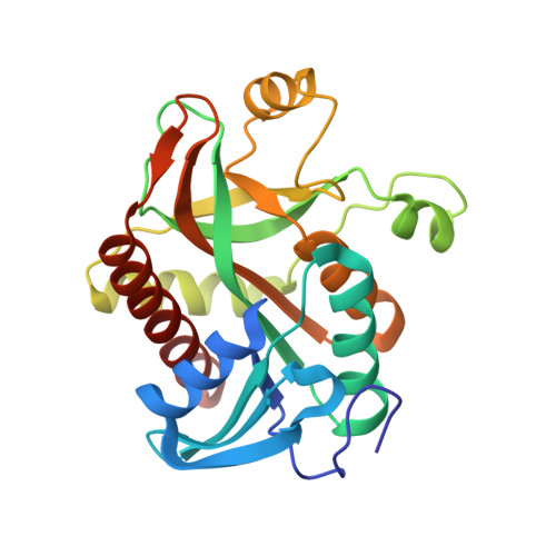
Crystal structure of E.coli purine nucleoside phosphorylase at 0.99 A resolution
Timofeev, V.I., Abramchik, Y.A., Esipov, R.S., Kuranova, I.P.To be published.
Experimental Data Snapshot
wwPDB Validation 3D Report Full Report

| Ligands 1 Unique | |||||
|---|---|---|---|---|---|
| ID | Chains | Name / Formula / InChI Key | 2D Diagram | 3D Interactions | |
| GOL Download:Ideal Coordinates CCD File | G [auth A] H [auth C] I [auth D] J [auth D] K [auth D] | GLYCEROL C3 H8 O3 PEDCQBHIVMGVHV-UHFFFAOYSA-N |  | ||
| Length ( Å ) | Angle ( ˚ ) |
|---|---|
| a = 74.12 | α = 90 |
| b = 110.22 | β = 111.08 |
| c = 88.21 | γ = 90 |
| Software Name | Purpose |
|---|---|
| HKL-2000 | data collection |
| PHASER | phasing |
| REFMAC | refinement |
| MOSFLM | data reduction |
| SCALA | data scaling |