4RGV

Crystal structure of the Methanocaldococcus jannaschii G1PDH


Experimental Data Snapshot

  • Method: X-RAY DIFFRACTION
  • Resolution: 2.45 Å
  • R-Value Free: 0.231 
  • R-Value Work: 0.193 
  • R-Value Observed: 0.193 

wwPDB Validation   3D Report Full Report


This is version 1.5 of the entry. See complete history


Literature

Structure and Evolution of the Archaeal Lipid Synthesis Enzyme sn-Glycerol-1-phosphate Dehydrogenase.

Carbone, V.Schofield, L.R.Zhang, Y.Sang, C.Dey, D.Hannus, I.M.Martin, W.F.Sutherland-Smith, A.J.Ronimus, R.S.

(2015) J Biol Chem 290: 21690-21704

  • DOI: https://doi.org/10.1074/jbc.M115.647461
  • Primary Citation of Related Structures:  
    4RFL, 4RGQ, 4RGV

  • PubMed Abstract: 

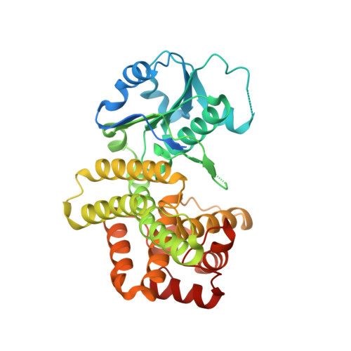
    One of the most critical events in the origins of cellular life was the development of lipid membranes. Archaea use isoprenoid chains linked via ether bonds to sn-glycerol 1-phosphate (G1P), whereas bacteria and eukaryotes use fatty acids attached via ester bonds to enantiomeric sn-glycerol 3-phosphate. NAD(P)H-dependent G1P dehydrogenase (G1PDH) forms G1P and has been proposed to have played a crucial role in the speciation of the Archaea. We present here, to our knowledge, the first structures of archaeal G1PDH from the hyperthermophilic methanogen Methanocaldococcus jannaschii with bound substrate dihydroxyacetone phosphate, product G1P, NADPH, and Zn(2+) cofactor. We also biochemically characterized the enzyme with respect to pH optimum, cation specificity, and kinetic parameters for dihydroxyacetone phosphate and NAD(P)H. The structures provide key evidence for the reaction mechanism in the stereospecific addition for the NAD(P)H-based pro-R hydrogen transfer and the coordination of the Zn(2+) cofactor during catalysis. Structure-based phylogenetic analyses also provide insight into the origins of G1PDH.


  • Organizational Affiliation

    From AgResearch Limited, Grasslands Research Centre, Tennent Drive, Private Bag 11008, Palmerston North 4442, New Zealand.


Macromolecules
Find similar proteins by:  (by identity cutoff)  |  3D Structure
Entity ID: 1
MoleculeChains Sequence LengthOrganismDetailsImage
Glycerol-1-phosphate dehydrogenase
A, B
368Methanocaldococcus jannaschii DSM 2661Mutation(s): 0 
Gene Names: egsAMJ0712
EC: 1.1.1.261
UniProt
Find proteins for Q58122 (Methanocaldococcus jannaschii (strain ATCC 43067 / DSM 2661 / JAL-1 / JCM 10045 / NBRC 100440))
Explore Q58122 
Go to UniProtKB:  Q58122
Entity Groups  
Sequence Clusters30% Identity50% Identity70% Identity90% Identity95% Identity100% Identity
UniProt GroupQ58122
Sequence Annotations
Expand
  • Reference Sequence
Experimental Data & Validation

Experimental Data

  • Method: X-RAY DIFFRACTION
  • Resolution: 2.45 Å
  • R-Value Free: 0.231 
  • R-Value Work: 0.193 
  • R-Value Observed: 0.193 
  • Space Group: P 1 21 1
Unit Cell:
Length ( Å )Angle ( ˚ )
a = 49.765α = 90
b = 59.552β = 90.39
c = 119.825γ = 90
Software Package:
Software NamePurpose
Blu-Icedata collection
PHASERphasing
REFMACrefinement
XDSdata reduction
SCALAdata scaling

Structure Validation

View Full Validation Report


Entry History 

Deposition Data

Revision History  (Full details and data files)

  • Version 1.0: 2015-07-22
    Type: Initial release
  • Version 1.1: 2015-09-02
    Changes: Database references
  • Version 1.2: 2015-09-23
    Changes: Database references
  • Version 1.3: 2015-09-30
    Changes: Data collection
  • Version 1.4: 2017-11-22
    Changes: Refinement description
  • Version 1.5: 2023-09-20
    Changes: Data collection, Database references, Derived calculations, Refinement description