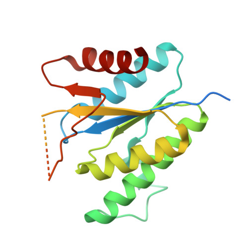
4RFV

Structure of the Mycobacterium tuberculosis APS kinase CysC Cys556Ala mutant


Experimental Data Snapshot

  • Method: X-RAY DIFFRACTION
  • Resolution: 1.69 Å
  • R-Value Free: 0.223 
  • R-Value Work: 0.184 
  • R-Value Observed: 0.186 

Starting Model: experimental
View more details

wwPDB Validation   3D Report Full Report


This is version 1.2 of the entry. See complete history


Literature

Crystal Structures of the Kinase Domain of the Sulfate-Activating Complex in Mycobacterium tuberculosis.

Poyraz, O.Brunner, K.Lohkamp, B.Axelsson, H.Hammarstrom, L.G.Schnell, R.Schneider, G.

(2015) PLoS One 10: e0121494-e0121494

  • DOI: https://doi.org/10.1371/journal.pone.0121494
  • Primary Citation of Related Structures:  
    4BZP, 4BZQ, 4BZX, 4RFV

  • PubMed Abstract: 

    In Mycobacterium tuberculosis the sulfate activating complex provides a key branching point in sulfate assimilation. The complex consists of two polypeptide chains, CysD and CysN. CysD is an ATP sulfurylase that, with the energy provided by the GTPase activity of CysN, forms adenosine-5'-phosphosulfate (APS) which can then enter the reductive branch of sulfate assimilation leading to the biosynthesis of cysteine. The CysN polypeptide chain also contains an APS kinase domain (CysC) that phosphorylates APS leading to 3'-phosphoadenosine-5'-phosphosulfate, the sulfate donor in the synthesis of sulfolipids. We have determined the crystal structures of CysC from M. tuberculosis as a binary complex with ADP, and as ternary complexes with ADP and APS and the ATP mimic AMP-PNP and APS, respectively, to resolutions of 1.5 Å, 2.1 Å and 1.7 Å, respectively. CysC shows the typical APS kinase fold, and the structures provide comprehensive views of the catalytic machinery, conserved in this enzyme family. Comparison to the structure of the human homolog show highly conserved APS and ATP binding sites, questioning the feasibility of the design of specific inhibitors of mycobacterial CysC. Residue Cys556 is part of the flexible lid region that closes off the active site upon substrate binding. Mutational analysis revealed this residue as one of the determinants controlling lid closure and hence binding of the nucleotide substrate.


  • Organizational Affiliation

    Department of Medical Biochemistry and Biophysics, Karolinska Institutet, Stockholm, Sweden.


Macromolecules
Find similar proteins by:  (by identity cutoff)  |  3D Structure
Entity ID: 1
MoleculeChains Sequence LengthOrganismDetailsImage
Bifunctional enzyme CysN/CysC
A, B
193Mycobacterium tuberculosis H37RvMutation(s): 1 
Gene Names: cysNcysNCMTCY373.05Rv1286
EC: 2.7.7.4 (PDB Primary Data), 2.7.1.25 (PDB Primary Data)
UniProt
Find proteins for P9WNM5 (Mycobacterium tuberculosis (strain ATCC 25618 / H37Rv))
Explore P9WNM5 
Go to UniProtKB:  P9WNM5
Entity Groups  
Sequence Clusters30% Identity50% Identity70% Identity90% Identity95% Identity100% Identity
UniProt GroupP9WNM5
Sequence Annotations
Expand
  • Reference Sequence
Small Molecules
Ligands 1 Unique
IDChains Name / Formula / InChI Key2D Diagram3D Interactions
PO4
Query on PO4

Download Ideal Coordinates CCD File 
C [auth A]PHOSPHATE ION
O4 P
NBIIXXVUZAFLBC-UHFFFAOYSA-K
Experimental Data & Validation

Experimental Data

  • Method: X-RAY DIFFRACTION
  • Resolution: 1.69 Å
  • R-Value Free: 0.223 
  • R-Value Work: 0.184 
  • R-Value Observed: 0.186 
  • Space Group: C 2 2 21
Unit Cell:
Length ( Å )Angle ( ˚ )
a = 68.167α = 90
b = 71.158β = 90
c = 118.56γ = 90
Software Package:
Software NamePurpose
DNAdata collection
PHASERphasing
REFMACrefinement
XDSdata reduction
Aimlessdata scaling

Structure Validation

View Full Validation Report


Entry History 

Deposition Data

Revision History  (Full details and data files)

  • Version 1.0: 2015-01-28
    Type: Initial release
  • Version 1.1: 2015-04-08
    Changes: Database references
  • Version 1.2: 2023-09-20
    Changes: Data collection, Database references, Derived calculations, Refinement description