4R7Y | pdb_00004r7y

Crystal structure of an active MCM hexamer


Experimental Data Snapshot

  • Method: X-RAY DIFFRACTION
  • Resolution: 2.70 Å
  • R-Value Free: 
    0.295 (Depositor), 0.295 (DCC) 
  • R-Value Work: 
    0.263 (Depositor), 0.263 (DCC) 
  • R-Value Observed: 
    0.263 (Depositor) 

Starting Models: experimental
View more details

wwPDB Validation 3D Report Full Report

Validation slider image for 4R7Y

Ligand Structure Quality Assessment 


This is version 1.5 of the entry. See complete history

Literature

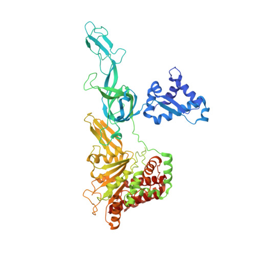
Analysis of the crystal structure of an active MCM hexamer.

Miller, J.M.Arachea, B.T.Epling, L.B.Enemark, E.J.

(2014) Elife 3: e03433-e03433

  • DOI: https://doi.org/10.7554/eLife.03433
  • Primary Citation Related Structures: 
    4R7Y, 4R7Z

  • PubMed Abstract: 

    In a previous Research article (Froelich et al., 2014), we suggested an MCM helicase activation mechanism, but were limited in discussing the ATPase domain because it was absent from the crystal structure. Here we present the crystal structure of a nearly full-length MCM hexamer that is helicase-active and thus has all features essential for unwinding DNA. The structure is a chimera of Sulfolobus solfataricus N-terminal domain and Pyrococcus furiosus ATPase domain. We discuss three major findings: 1) a novel conformation for the A-subdomain that could play a role in MCM regulation; 2) interaction of a universally conserved glutamine in the N-terminal Allosteric Communication Loop with the AAA+ domain helix-2-insert (h2i); and 3) a recessed binding pocket for the MCM ssDNA-binding motif influenced by the h2i. We suggest that during helicase activation, the h2i clamps down on the leading strand to facilitate strand retention and regulate ATP hydrolysis.


  • Organizational Affiliation
    • Department of Structural Biology, St Jude Children's Research Hospital, Memphis, United States.

Macromolecule Content 

  • Total Structure Weight: 140.19 kDa 
  • Atom Count: 9,494 
  • Modeled Residue Count: 1,182 
  • Deposited Residue Count: 1,226 
  • Unique protein chains: 1

Macromolecules

Find similar proteins by:|  3D Structure
Entity ID: 1
MoleculeChains  Sequence LengthOrganismDetailsImage
Minichromosome maintenance protein MCM, Cell division control protein 21
A, B
613Saccharolobus solfataricus P2Pyrococcus furiosus DSM 3638Mutation(s): 0 
Gene Names: MCMSSO0774PF0482
EC: 3.6.4.12 (PDB Primary Data), 5.6.2.4 (UniProt)
UniProt
Find proteins for Q8U3I4 (Pyrococcus furiosus (strain ATCC 43587 / DSM 3638 / JCM 8422 / Vc1))
Explore Q8U3I4 
Go to UniProtKB:  Q8U3I4
Find proteins for Q9UXG1 (Saccharolobus solfataricus (strain ATCC 35092 / DSM 1617 / JCM 11322 / P2))
Explore Q9UXG1 
Go to UniProtKB:  Q9UXG1
Entity Groups
Sequence Clusters30% Identity50% Identity70% Identity90% Identity95% Identity100% Identity
UniProt GroupsQ9UXG1Q8U3I4
Sequence Annotations
Expand
Reference Sequence

Small Molecules

Ligands 4 Unique
IDChains Name / Formula / InChI Key2D Diagram3D Interactions
ADP

Query on ADP


Download:Ideal Coordinates CCD File
C [auth A],
I [auth B]
ADENOSINE-5'-DIPHOSPHATE
C10 H15 N5 O10 P2
XTWYTFMLZFPYCI-KQYNXXCUSA-N
ZN

Query on ZN


Download:Ideal Coordinates CCD File
E [auth A],
K [auth B]
ZINC ION
Zn
PTFCDOFLOPIGGS-UHFFFAOYSA-N
CL

Query on CL


Download:Ideal Coordinates CCD File
F [auth A]
G [auth A]
H [auth A]
L [auth B]
M [auth B]
F [auth A],
G [auth A],
H [auth A],
L [auth B],
M [auth B],
N [auth B]
CHLORIDE ION
Cl
VEXZGXHMUGYJMC-UHFFFAOYSA-M
MG

Query on MG


Download:Ideal Coordinates CCD File
D [auth A],
J [auth B]
MAGNESIUM ION
Mg
JLVVSXFLKOJNIY-UHFFFAOYSA-N

Experimental Data & Validation

Experimental Data

  • Method: X-RAY DIFFRACTION
  • Resolution: 2.70 Å
  • R-Value Free:  0.295 (Depositor), 0.295 (DCC) 
  • R-Value Work:  0.263 (Depositor), 0.263 (DCC) 
  • R-Value Observed: 0.263 (Depositor) 
Space Group: P 63
Unit Cell:
Length ( Å )Angle ( ˚ )
a = 118.902α = 90
b = 118.902β = 90
c = 199.317γ = 120
Software Package:
Software NamePurpose
HKL-2000data collection
PHASERphasing
CNSrefinement
HKL-2000data reduction
HKL-2000data scaling

Structure Validation

View Full Validation Report


Ligand Structure Quality Assessment 


Entry History 

Deposition Data

Revision History  (Full details and data files)

  • Version 1.0: 2014-10-08
    Type: Initial release
  • Version 1.1: 2014-10-15
    Changes: Database references
  • Version 1.2: 2014-11-12
    Changes: Database references
  • Version 1.3: 2014-11-19
    Changes: Database references
  • Version 1.4: 2017-08-02
    Changes: Refinement description, Source and taxonomy
  • Version 1.5: 2023-09-20
    Changes: Data collection, Database references, Derived calculations, Refinement description