4R7G | pdb_00004r7g

Determination of the formylglycinamide ribonucleotide amidotransferase ammonia pathway by combining 3D-RISM theory with experiment


Experimental Data Snapshot

  • Method: X-RAY DIFFRACTION
  • Resolution: 2.90 Å
  • R-Value Free: 
    0.237 (Depositor), 0.243 (DCC) 
  • R-Value Work: 
    0.179 (Depositor), 0.185 (DCC) 

Starting Model: experimental
View more details

wwPDB Validation 3D Report Full Report

Validation slider image for 4R7G

Ligand Structure Quality Assessment 


This is version 1.3 of the entry. See complete history

Literature

Determination of the formylglycinamide ribonucleotide amidotransferase ammonia pathway by combining 3D-RISM theory with experiment.

Tanwar, A.S.Sindhikara, D.J.Hirata, F.Anand, R.

(2015) ACS Chem Biol 10: 698-704

  • DOI: https://doi.org/10.1021/cb501015r
  • Primary Citation Related Structures: 
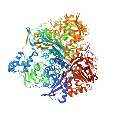
    4R7G

  • PubMed Abstract: 

    Molecular tunnels in enzyme systems possess variable architecture and are therefore difficult to predict. In this work, we design and apply an algorithm to resolve the pathway followed by ammonia using the bifunctional enzyme formylglycinamide ribonucleotide amidotransferase (FGAR-AT) as a model system. Though its crystal structure has been determined, an ammonia pathway connecting the glutaminase domain to the 30 Å distal FGAR/ATP binding site remains elusive. Crystallography suggested two purported paths: an N-terminal-adjacent path (path 1) and an auxiliary ADP-adjacent path (path 2). The algorithm presented here, RismPath, which enables fast and accurate determination of solvent distribution inside a protein channel, predicted path 2 as the preferred mode of ammonia transfer. Supporting experimental studies validate the identity of the path, and results lead to the conclusion that the residues in the middle of the channel do not partake in catalytic coupling and serve only as channel walls facilitating ammonia transfer.


  • Organizational Affiliation
    • †Department of Chemistry, Indian Institute of Technology, IIT-Bombay, Mumbai 400076, India.

Macromolecule Content 

  • Total Structure Weight: 143.66 kDa 
  • Atom Count: 10,182 
  • Modeled Residue Count: 1,284 
  • Deposited Residue Count: 1,303 
  • Unique protein chains: 1

Macromolecules

Find similar proteins by:|  3D Structure
Entity ID: 1
MoleculeChains  Sequence LengthOrganismDetailsImage
Phosphoribosylformylglycinamidine synthase1,303Salmonella enterica subsp. enterica serovar Typhimurium str. LT2Mutation(s): 1 
Gene Names: purLSTM2565
EC: 6.3.5.3
UniProt
Find proteins for P74881 (Salmonella typhimurium (strain LT2 / SGSC1412 / ATCC 700720))
Explore P74881 
Go to UniProtKB:  P74881
Entity Groups
Sequence Clusters30% Identity50% Identity70% Identity90% Identity95% Identity100% Identity
UniProt GroupP74881
Sequence Annotations
Expand
Reference Sequence

Small Molecules

Ligands 3 Unique
IDChains Name / Formula / InChI Key2D Diagram3D Interactions
ADP

Query on ADP


Download:Ideal Coordinates CCD File
K [auth A]ADENOSINE-5'-DIPHOSPHATE
C10 H15 N5 O10 P2
XTWYTFMLZFPYCI-KQYNXXCUSA-N
SO4

Query on SO4


Download:Ideal Coordinates CCD File
E [auth A]
F [auth A]
G [auth A]
H [auth A]
I [auth A]
E [auth A],
F [auth A],
G [auth A],
H [auth A],
I [auth A],
J [auth A]
SULFATE ION
O4 S
QAOWNCQODCNURD-UHFFFAOYSA-L
MG

Query on MG


Download:Ideal Coordinates CCD File
B [auth A],
C [auth A],
D [auth A]
MAGNESIUM ION
Mg
JLVVSXFLKOJNIY-UHFFFAOYSA-N
Modified Residues  1 Unique
IDChains TypeFormula2D DiagramParent
CYG
Query on CYG
A
L-PEPTIDE LINKINGC8 H14 N2 O5 SCYS

Experimental Data & Validation

Experimental Data

  • Method: X-RAY DIFFRACTION
  • Resolution: 2.90 Å
  • R-Value Free:  0.237 (Depositor), 0.243 (DCC) 
  • R-Value Work:  0.179 (Depositor), 0.185 (DCC) 
Space Group: P 65
Unit Cell:
Length ( Å )Angle ( ˚ )
a = 146.57α = 90
b = 146.57β = 90
c = 141.32γ = 120
Software Package:
Software NamePurpose
DENZOdata reduction
SCALEPACKdata scaling
CNSrefinement
PDB_EXTRACTdata extraction
HKL-2000data collection
HKL-2000data reduction
SCALAdata scaling
CNSphasing

Structure Validation

View Full Validation Report


Ligand Structure Quality Assessment 


Entry History 

Deposition Data

Revision History  (Full details and data files)

  • Version 1.0: 2015-01-21
    Type: Initial release
  • Version 1.1: 2017-11-22
    Changes: Refinement description
  • Version 1.2: 2022-08-24
    Changes: Database references, Derived calculations
  • Version 1.3: 2023-11-08
    Changes: Data collection, Refinement description