4R7E | pdb_00004r7e

Structure of Bre1 RING domain


Experimental Data Snapshot

  • Method: X-RAY DIFFRACTION
  • Resolution: 2.25 Å
  • R-Value Free: 
    0.247 (Depositor), 0.245 (DCC) 
  • R-Value Work: 
    0.221 (Depositor), 0.228 (DCC) 
  • R-Value Observed: 
    0.223 (Depositor) 

wwPDB Validation 3D Report Full Report

Validation slider image for 4R7E

This is version 1.2 of the entry. See complete history

Literature

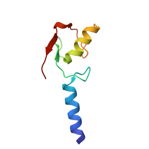
Structure of the yeast Bre1 RING domain.

Kumar, P.Wolberger, C.

(2015) Proteins 83: 1185-1190

  • DOI: https://doi.org/10.1002/prot.24812
  • Primary Citation Related Structures: 
    4R7E

  • PubMed Abstract: 

    Monoubiquitination of histone H2B at Lys123 in yeast plays a critical role in regulating transcription, mRNA export, DNA replication, and the DNA damage response. The RING E3 ligase, Bre1, catalyzes monoubiquitination of H2B in concert with the E2 ubiquitin-conjugating enzyme, Rad6. The crystal structure of a C-terminal fragment of Bre1 shows that the catalytic RING domain is preceded by an N-terminal helix that mediates coiled-coil interactions with a crystallographically related monomer. Homology modeling suggests that the human homologue of Bre1, RNF20/RNF40, heterodimerizes through similar coiled-coil interactions.


  • Organizational Affiliation
    • Department of Biophysics and Biophysical Chemistry, Johns Hopkins University School of Medicine, 725 North Wolfe Street, Baltimore, Maryland, 21205.

Macromolecule Content 

  • Total Structure Weight: 8.02 kDa 
  • Atom Count: 539 
  • Modeled Residue Count: 69 
  • Deposited Residue Count: 69 
  • Unique protein chains: 1

Macromolecules

Find similar proteins by:|  3D Structure
Entity ID: 1
MoleculeChains  Sequence LengthOrganismDetailsImage
E3 ubiquitin-protein ligase BRE169Saccharomyces cerevisiaeMutation(s): 0 
Gene Names: Bre1YDL074C
EC: 6.3.2 (PDB Primary Data), 2.3.2.27 (UniProt)
UniProt
Find proteins for Q07457 (Saccharomyces cerevisiae (strain ATCC 204508 / S288c))
Explore Q07457 
Go to UniProtKB:  Q07457
Entity Groups
Sequence Clusters30% Identity50% Identity70% Identity90% Identity95% Identity100% Identity
UniProt GroupQ07457
Sequence Annotations
Expand
Reference Sequence

Experimental Data & Validation

Experimental Data

  • Method: X-RAY DIFFRACTION
  • Resolution: 2.25 Å
  • R-Value Free:  0.247 (Depositor), 0.245 (DCC) 
  • R-Value Work:  0.221 (Depositor), 0.228 (DCC) 
  • R-Value Observed: 0.223 (Depositor) 
Space Group: P 61 2 2
Unit Cell:
Length ( Å )Angle ( ˚ )
a = 56.765α = 90
b = 56.765β = 90
c = 134.858γ = 120
Software Package:
Software NamePurpose
CBF:data collection
CBFlibdata collection
PHENIXmodel building
PHENIXrefinement
HKL-3000data reduction
HKL-3000data scaling
PHENIXphasing

Structure Validation

View Full Validation Report


Entry History 

Deposition Data

Revision History  (Full details and data files)

  • Version 1.0: 2015-05-13
    Type: Initial release
  • Version 1.1: 2015-08-26
    Changes: Database references
  • Version 1.2: 2024-02-28
    Changes: Data collection, Database references, Derived calculations