4R71

Structure of the Qbeta holoenzyme complex in the P1211 crystal form


Experimental Data Snapshot

  • Method: X-RAY DIFFRACTION
  • Resolution: 3.21 Å
  • R-Value Free: 0.268 
  • R-Value Work: 0.213 
  • R-Value Observed: 0.216 

wwPDB Validation   3D Report Full Report


This is version 1.2 of the entry. See complete history


Literature

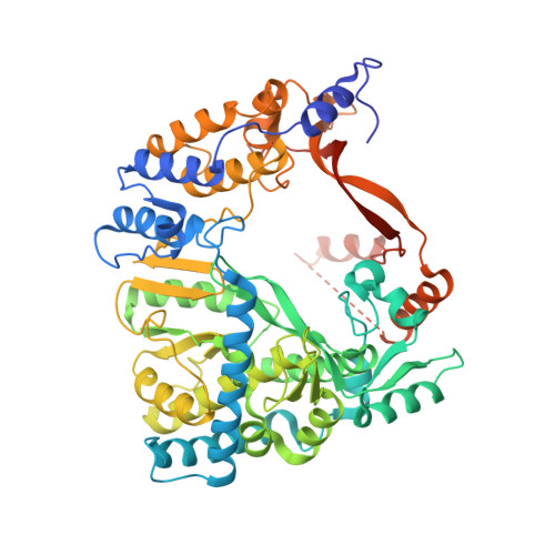
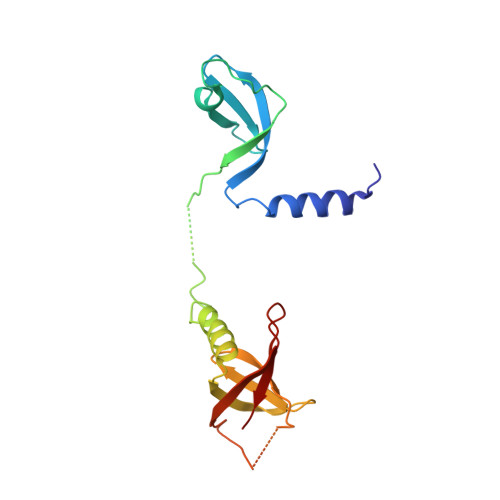
Structural basis for RNA-genome recognition during bacteriophage Q beta replication.

Gytz, H.Mohr, D.Seweryn, P.Yoshimura, Y.Kutlubaeva, Z.Dolman, F.Chelchessa, B.Chetverin, A.B.Mulder, F.A.Brodersen, D.E.Knudsen, C.R.

(2015) Nucleic Acids Res 43: 10893-10906

  • DOI: https://doi.org/10.1093/nar/gkv1212
  • Primary Citation of Related Structures:  
    4R71

  • PubMed Abstract: 

    Upon infection of Escherichia coli by bacteriophage Qβ, the virus-encoded β-subunit recruits host translation elongation factors EF-Tu and EF-Ts and ribosomal protein S1 to form the Qβ replicase holoenzyme complex, which is responsible for amplifying the Qβ (+)-RNA genome. Here, we use X-ray crystallography, NMR spectroscopy, as well as sequence conservation, surface electrostatic potential and mutational analyses to decipher the roles of the β-subunit and the first two oligonucleotide-oligosaccharide-binding domains of S1 (OB1-2) in the recognition of Qβ (+)-RNA by the Qβ replicase complex. We show how three basic residues of the β subunit form a patch located adjacent to the OB2 domain, and use NMR spectroscopy to demonstrate for the first time that OB2 is able to interact with RNA. Neutralization of the basic residues by mutagenesis results in a loss of both the phage infectivity in vivo and the ability of Qβ replicase to amplify the genomic RNA in vitro. In contrast, replication of smaller replicable RNAs is not affected. Taken together, our data suggest that the β-subunit and protein S1 cooperatively bind the (+)-stranded Qβ genome during replication initiation and provide a foundation for understanding template discrimination during replication initiation.


  • Organizational Affiliation

    Department of Molecular Biology and Genetics, Aarhus University, DK-8000 Aarhus C, Denmark.


Macromolecules
Find similar proteins by:  (by identity cutoff)  |  3D Structure
Entity ID: 1
MoleculeChains Sequence LengthOrganismDetailsImage
Elongation factor Ts, Elongation factor Tu
A, C
694Escherichia coli K-12Mutation(s): 0 
Gene Names: tsfb0170JW0165tufBb3980JW3943
UniProt
Find proteins for P0A6P1 (Escherichia coli (strain K12))
Explore P0A6P1 
Go to UniProtKB:  P0A6P1
Find proteins for P0CE48 (Escherichia coli (strain K12))
Explore P0CE48 
Go to UniProtKB:  P0CE48
Entity Groups  
Sequence Clusters30% Identity50% Identity70% Identity90% Identity95% Identity100% Identity
UniProt GroupsP0A6P1P0CE48
Sequence Annotations
Expand
  • Reference Sequence
Find similar proteins by:  (by identity cutoff)  |  3D Structure
Entity ID: 2
MoleculeChains Sequence LengthOrganismDetailsImage
RNA-directed RNA polymerase beta chain
B, D
595Qubevirus durumMutation(s): 0 
EC: 2.7.7.48
UniProt
Find proteins for P14647 (Escherichia virus Qbeta)
Explore P14647 
Go to UniProtKB:  P14647
Entity Groups  
Sequence Clusters30% Identity50% Identity70% Identity90% Identity95% Identity100% Identity
UniProt GroupP14647
Sequence Annotations
Expand
  • Reference Sequence
Find similar proteins by:  (by identity cutoff)  |  3D Structure
Entity ID: 3
MoleculeChains Sequence LengthOrganismDetailsImage
30S ribosomal protein S1
E, F
171Escherichia coli K-12Mutation(s): 0 
Gene Names: rpsAssyFb0911JW0894
UniProt
Find proteins for P0AG67 (Escherichia coli (strain K12))
Explore P0AG67 
Go to UniProtKB:  P0AG67
Entity Groups  
Sequence Clusters30% Identity50% Identity70% Identity90% Identity95% Identity100% Identity
UniProt GroupP0AG67
Sequence Annotations
Expand
  • Reference Sequence
Experimental Data & Validation

Experimental Data

  • Method: X-RAY DIFFRACTION
  • Resolution: 3.21 Å
  • R-Value Free: 0.268 
  • R-Value Work: 0.213 
  • R-Value Observed: 0.216 
  • Space Group: P 1 21 1
Unit Cell:
Length ( Å )Angle ( ˚ )
a = 99.73α = 90
b = 115.45β = 96.14
c = 178.52γ = 90
Software Package:
Software NamePurpose
PXIIIdata collection
PHASERphasing
PHENIXrefinement
XDSdata reduction
XSCALEdata scaling

Structure Validation

View Full Validation Report


Entry History 

Deposition Data

Revision History  (Full details and data files)

  • Version 1.0: 2015-09-23
    Type: Initial release
  • Version 1.1: 2017-06-07
    Changes: Database references
  • Version 1.2: 2024-02-28
    Changes: Data collection, Database references, Refinement description