4R1N | pdb_00004r1n

Crystal structure of (S)-3-hydroxybutylryl-CoA dehydrogenase form the n-butanol sysnthesizing bacterium, Clostridium butyricum.


Experimental Data Snapshot

  • Method: X-RAY DIFFRACTION
  • Resolution: 1.80 Å
  • R-Value Free: 
    0.240 (Depositor), 0.247 (DCC) 
  • R-Value Work: 
    0.207 (Depositor), 0.215 (DCC) 
  • R-Value Observed: 
    0.208 (Depositor) 

Starting Model: experimental
View more details

wwPDB Validation 3D Report Full Report

Validation slider image for 4R1N

This is version 1.1 of the entry. See complete history

Literature

Crystal structure of (S)-3-hydroxybutyryl-CoA dehydrogenase from Clostridium butyricum and its mutations that enhance reaction kinetics

Kim, E.J.Kim, J.Ahn, J.W.Kim, Y.J.Chang, J.H.Kim, K.J.

(2014) J Microbiol Biotechnol 24: 1636-1643

  • DOI: https://doi.org/10.4014/jmb.1407.07027
  • Primary Citation Related Structures: 
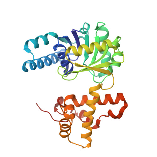
    4R1N

  • PubMed Abstract: 

    3-Hydroxybutyryl-CoA dehydrogenase is an enzyme that catalyzes the second step in the biosynthesis of n-butanol from acetyl-CoA, in which acetoacetyl-CoA is reduced to 3-hydroxybutyryl-CoA. To understand the molecular mechanisms of n-butanol biosynthesis, we determined the crystal structure of 3-hydroxybutyryl-CoA dehydrogenase from Clostridium butyricum (CbHBD). The monomer structure of CbHBD exhibits a two-domain topology, with N- and C-terminal domains, and the dimerization of the enzyme was mostly constituted at the C-terminal domain. The mode of cofactor binding to CbHBD was elucidated by determining the crystal structure of the enzyme in complex with NAD(+). We also determined the enzyme's structure in complex with its acetoacetyl-CoA substrate, revealing that the adenosine diphosphate moiety was not highly stabilized compared with the remainder of the acetoacetyl-CoA molecule. Using this structural information, we performed a series of sitedirected mutagenesis experiments on the enzyme, such as changing residues located near the substrate-binding site, and finally developed a highly efficient CbHBD K50A/K54A/L232Y triple mutant enzyme that exhibited approximately 5-fold higher enzyme activity than did the wild type. The increased enzyme activity of the mutant was confirmed by enzyme kinetic measurements. The highly efficient mutant enzyme should be useful for increasing the production rate of n-butanol.


  • Organizational Affiliation
    • School of Life Sciences, KNU Creative BioResearch Group, Kyungpook National University, Daegu 702-701, Republic of Korea.

Macromolecule Content 

  • Total Structure Weight: 121.98 kDa 
  • Atom Count: 8,756 
  • Modeled Residue Count: 1,128 
  • Deposited Residue Count: 1,128 
  • Unique protein chains: 1

Macromolecules

Find similar proteins by:|  3D Structure
Entity ID: 1
MoleculeChains  Sequence LengthOrganismDetailsImage
3-hydroxybutyryl-CoA dehydrogenase
A, B, C, D
282Clostridium butyricum E4 str. BoNT E BL5262Mutation(s): 0 
Gene Names: CLP_3850hbd
EC: 1.1.1.157
UniProt
Find proteins for C4IEM5 (Clostridium butyricum E4 str. BoNT E BL5262)
Explore C4IEM5 
Go to UniProtKB:  C4IEM5
Entity Groups
Sequence Clusters30% Identity50% Identity70% Identity90% Identity95% Identity100% Identity
UniProt GroupC4IEM5
Sequence Annotations
Expand
Reference Sequence

Experimental Data & Validation

Experimental Data

  • Method: X-RAY DIFFRACTION
  • Resolution: 1.80 Å
  • R-Value Free:  0.240 (Depositor), 0.247 (DCC) 
  • R-Value Work:  0.207 (Depositor), 0.215 (DCC) 
  • R-Value Observed: 0.208 (Depositor) 
Space Group: H 3
Unit Cell:
Length ( Å )Angle ( ˚ )
a = 146.475α = 90
b = 146.475β = 90
c = 202.295γ = 120
Software Package:
Software NamePurpose
REFMACrefinement
PDB_EXTRACTdata extraction
HKL-2000data collection
HKL-2000data reduction
HKL-2000data scaling

Structure Validation

View Full Validation Report


Entry History 

Deposition Data

Revision History  (Full details and data files)

  • Version 1.0: 2015-07-29
    Type: Initial release
  • Version 1.1: 2023-11-08
    Changes: Data collection, Database references, Refinement description