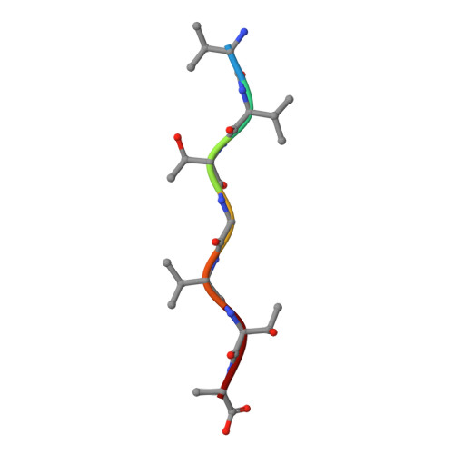
4R0W

Vvtgvta, an amyloid forming segment from alpha synuclein, residues 70-76


Experimental Data Snapshot

  • Method: X-RAY DIFFRACTION
  • Resolution: 1.50 Å
  • R-Value Free: 0.274 
  • R-Value Work: 0.244 
  • R-Value Observed: 0.247 

Starting Model: in silico
View more details

wwPDB Validation   3D Report Full Report


This is version 1.3 of the entry. See complete history


Literature

Structure-based design of functional amyloid materials.

Li, D.Jones, E.M.Sawaya, M.R.Furukawa, H.Luo, F.Ivanova, M.Sievers, S.A.Wang, W.Yaghi, O.M.Liu, C.Eisenberg, D.S.

(2014) J Am Chem Soc 136: 18044-18051

  • DOI: https://doi.org/10.1021/ja509648u
  • Primary Citation of Related Structures:  
    4R0P, 4R0U, 4R0W

  • PubMed Abstract: 

    Amyloid fibers, once exclusively associated with disease, are acquiring utility as a class of biological nanomaterials. Here we introduce a method that utilizes the atomic structures of amyloid peptides, to design materials with versatile applications. As a model application, we designed amyloid fibers capable of capturing carbon dioxide from flue gas, to address the global problem of excess anthropogenic carbon dioxide. By measuring dynamic separation of carbon dioxide from nitrogen, we show that fibers with designed amino acid sequences double the carbon dioxide binding capacity of the previously reported fiber formed by VQIVYK from Tau protein. In a second application, we designed fibers that facilitate retroviral gene transfer. By measuring lentiviral transduction, we show that designed fibers exceed the efficiency of polybrene, a commonly used enhancer of transduction. The same procedures can be adapted to the design of countless other amyloid materials with a variety of properties and uses.


  • Organizational Affiliation

    UCLA-DOE Institute, and Department of Chemistry and Biochemistry, University of California , Los Angeles, California 90095, United States.


Macromolecules

Find similar proteins by:  Sequence   |   3D Structure  

Entity ID: 1
MoleculeChains Sequence LengthOrganismDetailsImage
Alpha-synuclein7Homo sapiensMutation(s): 0 
UniProt & NIH Common Fund Data Resources
Find proteins for P37840 (Homo sapiens)
Explore P37840 
Go to UniProtKB:  P37840
PHAROS:  P37840
GTEx:  ENSG00000145335 
Entity Groups  
UniProt GroupP37840
Sequence Annotations
Expand
  • Reference Sequence
Experimental Data & Validation

Experimental Data

  • Method: X-RAY DIFFRACTION
  • Resolution: 1.50 Å
  • R-Value Free: 0.274 
  • R-Value Work: 0.244 
  • R-Value Observed: 0.247 
  • Space Group: C 1 2 1
Unit Cell:
Length ( Å )Angle ( ˚ )
a = 50.367α = 90
b = 4.823β = 107.89
c = 15.692γ = 90
Software Package:
Software NamePurpose
DENZOdata reduction
SCALEPACKdata scaling
PHASERphasing
REFMACrefinement
PDB_EXTRACTdata extraction

Structure Validation

View Full Validation Report


Entry History 

Deposition Data

Revision History  (Full details and data files)

  • Version 1.0: 2014-12-17
    Type: Initial release
  • Version 1.1: 2015-01-14
    Changes: Database references
  • Version 1.2: 2024-02-28
    Changes: Data collection, Database references
  • Version 1.3: 2024-04-03
    Changes: Refinement description