4QX5

Neutron diffraction reveals hydrogen bonds critical for cGMP-selective activation: Insights for PKG agonist design


Experimental Data Snapshot

  • Method: X-RAY DIFFRACTION
  • Resolution: 1.32 Å
  • R-Value Free: 0.224 
  • R-Value Work: 0.202 
  • R-Value Observed: 0.203 

wwPDB Validation   3D Report Full Report


Ligand Structure Quality Assessment 


This is version 1.2 of the entry. See complete history


Literature

Neutron Diffraction Reveals Hydrogen Bonds Critical for cGMP-Selective Activation: Insights for cGMP-Dependent Protein Kinase Agonist Design.

Huang, G.Y.Gerlits, O.O.Blakeley, M.P.Sankaran, B.Kovalevsky, A.Y.Kim, C.

(2014) Biochemistry 53: 6725-6727

  • DOI: https://doi.org/10.1021/bi501012v
  • Primary Citation of Related Structures:  
    4QX5, 4QXK

  • PubMed Abstract: 

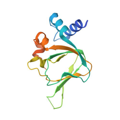
    High selectivity of cyclic-nucleotide binding (CNB) domains for cAMP and cGMP are required for segregating signaling pathways; however, the mechanism of selectivity remains unclear. To investigate the mechanism of high selectivity in cGMP-dependent protein kinase (PKG), we determined a room-temperature joint X-ray/neutron (XN) structure of PKG Iβ CNB-B, a domain 200-fold selective for cGMP over cAMP, bound to cGMP (2.2 Å), and a low-temperature X-ray structure of CNB-B with cAMP (1.3 Å). The XN structure directly describes the hydrogen bonding interactions that modulate high selectivity for cGMP, while the structure with cAMP reveals that all these contacts are disrupted, explaining its low affinity for cAMP.


  • Organizational Affiliation

    Verna and Mars McClean Department of Biochemistry and Molecular Biology, Baylor College of Medicine , One Baylor Plaza, Houston, Texas 77004, United States.


Macromolecules
Find similar proteins by:  (by identity cutoff)  |  3D Structure
Entity ID: 1
MoleculeChains Sequence LengthOrganismDetailsImage
cGMP-dependent protein kinase 1153Homo sapiensMutation(s): 0 
Gene Names: PRKG1PRKG1BPRKGR1APRKGR1B
EC: 2.7.11.12
UniProt & NIH Common Fund Data Resources
Find proteins for Q13976 (Homo sapiens)
Explore Q13976 
Go to UniProtKB:  Q13976
PHAROS:  Q13976
GTEx:  ENSG00000185532 
Entity Groups  
Sequence Clusters30% Identity50% Identity70% Identity90% Identity95% Identity100% Identity
UniProt GroupQ13976
Sequence Annotations
Expand
  • Reference Sequence
Small Molecules
Ligands 2 Unique
IDChains Name / Formula / InChI Key2D Diagram3D Interactions
CMP
Query on CMP

Download Ideal Coordinates CCD File 
B [auth A]ADENOSINE-3',5'-CYCLIC-MONOPHOSPHATE
C10 H12 N5 O6 P
IVOMOUWHDPKRLL-KQYNXXCUSA-N
IOD
Query on IOD

Download Ideal Coordinates CCD File 
C [auth A]
D [auth A]
E [auth A]
F [auth A]
G [auth A]
C [auth A],
D [auth A],
E [auth A],
F [auth A],
G [auth A],
H [auth A],
I [auth A],
J [auth A],
K [auth A],
L [auth A],
M [auth A],
N [auth A],
O [auth A],
P [auth A],
Q [auth A],
R [auth A],
S [auth A],
T [auth A]
IODIDE ION
I
XMBWDFGMSWQBCA-UHFFFAOYSA-M
Experimental Data & Validation

Experimental Data

  • Method: X-RAY DIFFRACTION
  • Resolution: 1.32 Å
  • R-Value Free: 0.224 
  • R-Value Work: 0.202 
  • R-Value Observed: 0.203 
  • Space Group: P 41 21 2
Unit Cell:
Length ( Å )Angle ( ˚ )
a = 48.44α = 90
b = 48.44β = 90
c = 103.462γ = 90
Software Package:
Software NamePurpose
Specdata collection
PHENIXmodel building
PHENIXrefinement
HKL-2000data reduction
HKL-2000data scaling
PHENIXphasing

Structure Validation

View Full Validation Report


Ligand Structure Quality Assessment 


Entry History 

Deposition Data

Revision History  (Full details and data files)

  • Version 1.0: 2014-11-12
    Type: Initial release
  • Version 1.1: 2017-11-22
    Changes: Refinement description
  • Version 1.2: 2024-02-28
    Changes: Data collection, Database references, Derived calculations