4QWQ | pdb_00004qwq

Crystal structure of the DNA-binding domain of the response regulator SaeR from Staphylococcus aureus


Experimental Data Snapshot

  • Method: X-RAY DIFFRACTION
  • Resolution: 2.50 Å
  • R-Value Free: 
    0.281 (Depositor), 0.273 (DCC) 
  • R-Value Work: 
    0.205 (Depositor), 0.234 (DCC) 
  • R-Value Observed: 
    0.209 (Depositor) 

wwPDB Validation 3D Report Full Report

Validation slider image for 4QWQ

This is version 1.1 of the entry. See complete history

Literature

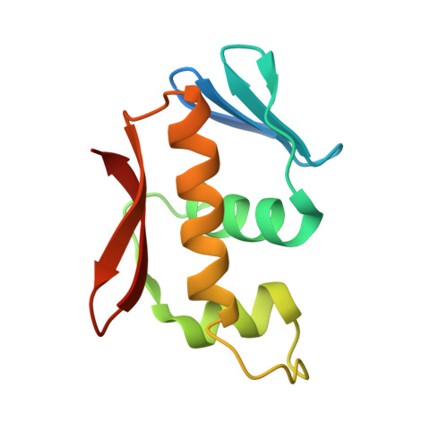
Structure of the DNA-binding domain of the response regulator SaeR from Staphylococcus aureus.

Fan, X.Zhang, X.Zhu, Y.Niu, L.Teng, M.Sun, B.Li, X.

(2015) Acta Crystallogr D Biol Crystallogr 71: 1768-1776

  • DOI: https://doi.org/10.1107/S1399004715010287
  • Primary Citation Related Structures: 
    4QWQ

  • PubMed Abstract: 

    The SaeR/S two-component regulatory system is essential for controlling the expression of many virulence factors in Staphylococcus aureus. SaeR, a member of the OmpR/PhoB family, is a response regulator with an N-terminal regulatory domain and a C-terminal DNA-binding domain. In order to elucidate how SaeR binds to the promoter regions of target genes, the crystal structure of the DNA-binding domain of SaeR (SaeR(DBD)) was solved at 2.5 Å resolution. The structure reveals that SaeR(DBD) exists as a monomer and has the canonical winged helix-turn-helix module. EMSA experiments suggested that full-length SaeR can bind to the P1 promoter and that the binding affinity is higher than that of its C-terminal DNA-binding domain. Five key residues on the winged helix-turn-helix module were verified to be important for binding to the P1 promoter in vitro and for the physiological function of SaeR in vivo.


  • Organizational Affiliation
    • Hefei National Laboratory for Physical Sciences at Microscale and School of Life Sciences, University of Science and Technology of China, Hefei, Anhui 230026, People's Republic of China.

Macromolecule Content 

  • Total Structure Weight: 26.49 kDa 
  • Atom Count: 1,585 
  • Modeled Residue Count: 196 
  • Deposited Residue Count: 220 
  • Unique protein chains: 1

Macromolecules

Find similar proteins by:|  3D Structure
Entity ID: 1
MoleculeChains  Sequence LengthOrganismDetailsImage
Response regulator SaeRA [auth B],
B [auth A]
110Staphylococcus aureusMutation(s): 0 
Gene Names: saeRCH52_02235X998_0755
UniProt
Find proteins for Q2G2G2 (Staphylococcus aureus (strain NCTC 8325 / PS 47))
Explore Q2G2G2 
Go to UniProtKB:  Q2G2G2
Entity Groups
Sequence Clusters30% Identity50% Identity70% Identity90% Identity95% Identity100% Identity
UniProt GroupQ2G2G2
Sequence Annotations
Expand
Reference Sequence

Experimental Data & Validation

Experimental Data

  • Method: X-RAY DIFFRACTION
  • Resolution: 2.50 Å
  • R-Value Free:  0.281 (Depositor), 0.273 (DCC) 
  • R-Value Work:  0.205 (Depositor), 0.234 (DCC) 
  • R-Value Observed: 0.209 (Depositor) 
Space Group: P 21 21 21
Unit Cell:
Length ( Å )Angle ( ˚ )
a = 31.974α = 90
b = 62.648β = 90
c = 116.262γ = 90
Software Package:
Software NamePurpose
HKL-2000data collection
PHENIXrefinement
HKL-2000data reduction
HKL-2000data scaling

Structure Validation

View Full Validation Report


Entry History 

Deposition Data

Revision History  (Full details and data files)

  • Version 1.0: 2015-08-19
    Type: Initial release
  • Version 1.1: 2024-03-20
    Changes: Data collection, Database references