


Crystal structure of predicted N-acyltransferase (ypeA) in complex with acetyl-CoA from Escherichia coli
Filippova, E.V., Minasov, G., Winsor, G., Dubrovska, I., Shuvalova, L., Wolfe, A.J., Anderson, W.F.To be published.
Experimental Data Snapshot
Starting Model: experimental
View more details
Entity ID: 1 | |||||
|---|---|---|---|---|---|
| Molecule | Chains | Sequence Length | Organism | Details | Image |
| Acetyltransferase YpeA | 158 | Escherichia coli K-12 | Mutation(s): 0 Gene Names: b2434, JW2427, ypeA EC: 2.3.1 | 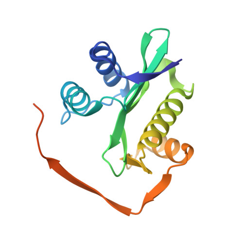 | |
UniProt | |||||
Entity Groups | |||||
| Sequence Clusters | 30% Identity50% Identity70% Identity90% Identity95% Identity100% Identity | ||||
| UniProt Group | P76539 | ||||
Sequence AnnotationsExpand | |||||
Reference Sequence | |||||
| Ligands 2 Unique | |||||
|---|---|---|---|---|---|
| ID | Chains | Name / Formula / InChI Key | 2D Diagram | 3D Interactions | |
| ACO Download:Ideal Coordinates CCD File | I [auth A] L [auth B] N [auth C] P [auth D] R [auth E] | ACETYL COENZYME *A C23 H38 N7 O17 P3 S ZSLZBFCDCINBPY-ZSJPKINUSA-N |  | ||
| SO4 Download:Ideal Coordinates CCD File | J [auth A] K [auth A] M [auth B] O [auth C] Q [auth D] | SULFATE ION O4 S QAOWNCQODCNURD-UHFFFAOYSA-L |  | ||
| Length ( Å ) | Angle ( ˚ ) |
|---|---|
| a = 67.334 | α = 90 |
| b = 139.968 | β = 112.11 |
| c = 75.358 | γ = 90 |
| Software Name | Purpose |
|---|---|
| Blu-Ice | data collection |
| PHASER | phasing |
| REFMAC | refinement |
| HKL-3000 | data reduction |
| HKL-3000 | data scaling |


