4QVB | pdb_00004qvb

Mycobacterium tuberculosis protein Rv1155 in complex with co-enzyme F420


Experimental Data Snapshot

  • Method: X-RAY DIFFRACTION
  • Resolution: 2.30 Å
  • R-Value Free: 
    0.254 (Depositor), 0.269 (DCC) 
  • R-Value Work: 
    0.183 (Depositor) 
  • R-Value Observed: 
    0.187 (Depositor) 

wwPDB Validation 3D Report Full Report

Validation slider image for 4QVB

Ligand Structure Quality Assessment 


This is version 1.2 of the entry. See complete history

Literature

Molecular insights into the binding of coenzyme F420 to the conserved protein Rv1155 from Mycobacterium tuberculosis.

Mashalidis, E.H.Gittis, A.G.Tomczak, A.Abell, C.Barry, C.E.Garboczi, D.N.

(2015) Protein Sci 24: 729-740

  • DOI: https://doi.org/10.1002/pro.2645
  • Primary Citation Related Structures: 
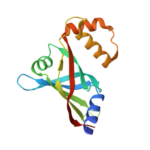
    4QVB

  • PubMed Abstract: 

    Coenzyme F420 is a deazaflavin hydride carrier with a lower reduction potential than most flavins. In Mycobacterium tuberculosis (Mtb), F420 plays an important role in activating PA-824, an antituberculosis drug currently used in clinical trials. Although F420 is important to Mtb redox metabolism, little is known about the enzymes that bind F420 and the reactions that they catalyze. We have identified a novel F420 -binding protein, Rv1155, which is annotated in the Mtb genome sequence as a putative flavin mononucleotide (FMN)-binding protein. Using biophysical techniques, we have demonstrated that instead of binding FMN or other flavins, Rv1155 binds coenzyme F420 . The crystal structure of the complex of Rv1155 and F420 reveals one F420 molecule bound to each monomer of the Rv1155 dimer. Structural, biophysical, and bioinformatic analyses of the Rv1155-F420 complex provide clues about its role in the bacterium.


  • Organizational Affiliation
    • Tuberculosis Research Section, National Institute of Allergy and Infectious Diseases, Bethesda, Maryland, 20892; Department of Chemistry, University of Cambridge, Cambridge, CB2 1EW, United Kingdom.

Macromolecule Content 

  • Total Structure Weight: 34.92 kDa 
  • Atom Count: 2,400 
  • Modeled Residue Count: 285 
  • Deposited Residue Count: 294 
  • Unique protein chains: 1

Macromolecules

Find similar proteins by:|  3D Structure
Entity ID: 1
MoleculeChains  Sequence LengthOrganismDetailsImage
Rv1155 protein
A, B
147Mycobacterium tuberculosis 7199-99Mutation(s): 0 
Gene Names: MT7199_1184
EC: 1
UniProt
Find proteins for O06553 (Mycobacterium tuberculosis (strain ATCC 25618 / H37Rv))
Explore O06553 
Go to UniProtKB:  O06553
Entity Groups
Sequence Clusters30% Identity50% Identity70% Identity90% Identity95% Identity100% Identity
UniProt GroupO06553
Sequence Annotations
Expand
Reference Sequence

Small Molecules

Ligands 6 Unique
IDChains Name / Formula / InChI Key2D Diagram3D Interactions
F42

Query on F42


Download:Ideal Coordinates CCD File
M [auth B],
N [auth B]
COENZYME F420
C29 H36 N5 O18 P
GEHSZWRGPHDXJO-NALJQGANSA-N
PGO

Query on PGO


Download:Ideal Coordinates CCD File
L [auth A]S-1,2-PROPANEDIOL
C3 H8 O2
DNIAPMSPPWPWGF-VKHMYHEASA-N
PDO

Query on PDO


Download:Ideal Coordinates CCD File
F [auth A],
G [auth A],
Q [auth B],
R [auth B]
1,3-PROPANDIOL
C3 H8 O2
YPFDHNVEDLHUCE-UHFFFAOYSA-N
EDO

Query on EDO


Download:Ideal Coordinates CCD File
H [auth A],
I [auth A],
J [auth A],
K [auth A]
1,2-ETHANEDIOL
C2 H6 O2
LYCAIKOWRPUZTN-UHFFFAOYSA-N
FMT

Query on FMT


Download:Ideal Coordinates CCD File
C [auth A],
D [auth A],
E [auth A],
O [auth B],
P [auth B]
FORMIC ACID
C H2 O2
BDAGIHXWWSANSR-UHFFFAOYSA-N
NA

Query on NA


Download:Ideal Coordinates CCD File
S [auth B]SODIUM ION
Na
FKNQFGJONOIPTF-UHFFFAOYSA-N

Experimental Data & Validation

Experimental Data

  • Method: X-RAY DIFFRACTION
  • Resolution: 2.30 Å
  • R-Value Free:  0.254 (Depositor), 0.269 (DCC) 
  • R-Value Work:  0.183 (Depositor) 
  • R-Value Observed: 0.187 (Depositor) 
Space Group: P 21 21 21
Unit Cell:
Length ( Å )Angle ( ˚ )
a = 53.579α = 90
b = 65.172β = 90
c = 77.063γ = 90
Software Package:
Software NamePurpose
SERGUIdata collection
MOLREPphasing
PHENIXrefinement
XDSdata reduction
XDSdata scaling

Structure Validation

View Full Validation Report


Ligand Structure Quality Assessment 


Entry History 

Deposition Data

Revision History  (Full details and data files)

  • Version 1.0: 2015-02-25
    Type: Initial release
  • Version 1.1: 2015-05-20
    Changes: Database references
  • Version 1.2: 2024-02-28
    Changes: Data collection, Database references, Derived calculations