4QQH

Crystal structure of C1QL3 in space group H32


Experimental Data Snapshot

  • Method: X-RAY DIFFRACTION
  • Resolution: 1.20 Å
  • R-Value Free: 0.167 
  • R-Value Work: 0.154 
  • R-Value Observed: 0.154 

wwPDB Validation   3D Report Full Report


This is version 1.3 of the entry. See complete history


Literature

Structures of C1q-like Proteins Reveal Unique Features among the C1q/TNF Superfamily.

Ressl, S.Vu, B.K.Vivona, S.Martinelli, D.C.Sudhof, T.C.Brunger, A.T.

(2015) Structure 23: 688-699

  • DOI: https://doi.org/10.1016/j.str.2015.01.019
  • Primary Citation of Related Structures:  
    4QPY, 4QQ2, 4QQH, 4QQL, 4QQO, 4QQP

  • PubMed Abstract: 

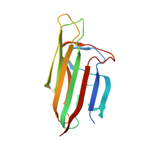
    C1q-like (C1QL) -1, -2, and -3 proteins are encoded by homologous genes that are highly expressed in brain. C1QLs bind to brain-specific angiogenesis inhibitor 3 (BAI3), an adhesion-type G-protein coupled receptor that may regulate dendritic morphology by organizing actin filaments. To begin to understand the function of C1QLs, we determined high-resolution crystal structures of the globular C1q-domains of C1QL1, C1QL2, and C1QL3. Each structure is a trimer, with each protomer forming a jelly-roll fold consisting of 10 β strands. Moreover, C1QL trimers may assemble into higher-order oligomers similar to adiponectin and contain four Ca(2+)-binding sites along the trimeric symmetry axis, as well as additional surface Ca(2+)-binding sites. Mutation of Ca(2+)-coordinating residues along the trimeric symmetry axis lowered the Ca(2+)-binding affinity and protein stability. Our results reveal unique structural features of C1QLs among C1q/TNF superfamily proteins that may be associated with their specific brain functions.


  • Organizational Affiliation

    Department of Molecular and Cellular Physiology, Stanford University, Stanford, CA 94305, USA. Electronic address: suressl@indiana.edu.


Macromolecules
Find similar proteins by:  (by identity cutoff)  |  3D Structure
Entity ID: 1
MoleculeChains Sequence LengthOrganismDetailsImage
Complement C1q-like protein 3137Mus musculusMutation(s): 0 
Gene Names: C1ql3C1qlCtrp13
UniProt & NIH Common Fund Data Resources
Find proteins for Q9ESN4 (Mus musculus)
Explore Q9ESN4 
Go to UniProtKB:  Q9ESN4
IMPC:  MGI:2387350
Entity Groups  
Sequence Clusters30% Identity50% Identity70% Identity90% Identity95% Identity100% Identity
UniProt GroupQ9ESN4
Sequence Annotations
Expand
  • Reference Sequence
Experimental Data & Validation

Experimental Data

  • Method: X-RAY DIFFRACTION
  • Resolution: 1.20 Å
  • R-Value Free: 0.167 
  • R-Value Work: 0.154 
  • R-Value Observed: 0.154 
  • Space Group: H 3 2
Unit Cell:
Length ( Å )Angle ( ˚ )
a = 76.23α = 90
b = 76.23β = 90
c = 125.407γ = 120
Software Package:
Software NamePurpose
Blu-Icedata collection
PHASERphasing
PHENIXrefinement
XDSdata reduction
SCALAdata scaling

Structure Validation

View Full Validation Report


Entry History 

Deposition Data

Revision History  (Full details and data files)

  • Version 1.0: 2015-04-15
    Type: Initial release
  • Version 1.1: 2015-05-06
    Changes: Database references
  • Version 1.2: 2017-11-22
    Changes: Refinement description
  • Version 1.3: 2024-02-28
    Changes: Data collection, Database references, Derived calculations