4QNL | pdb_00004qnl

Crystal structure of tail fiber protein gp63.1 from E. coli phage G7C


Experimental Data Snapshot

  • Method: X-RAY DIFFRACTION
  • Resolution: 2.41 Å
  • R-Value Free: 
    0.221 (Depositor), 0.221 (DCC) 
  • R-Value Work: 
    0.159 (Depositor), 0.164 (DCC) 
  • R-Value Observed: 
    0.162 (Depositor) 

wwPDB Validation 3D Report Full Report

Validation slider image for 4QNL

This is version 1.4 of the entry. See complete history

Literature

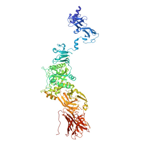
Function of bacteriophage G7C esterase tailspike in host cell adsorption.

Prokhorov, N.S.Riccio, C.Zdorovenko, E.L.Shneider, M.M.Browning, C.Knirel, Y.A.Leiman, P.G.Letarov, A.V.

(2017) Mol Microbiol 105: 385-398

  • DOI: https://doi.org/10.1111/mmi.13710
  • Primary Citation Related Structures: 
    4QNL

  • PubMed Abstract: 

    Bacteriophages recognize and bind to their hosts with the help of receptor-binding proteins (RBPs) that emanate from the phage particle in the form of fibers or tailspikes. RBPs show a great variability in their shapes, sizes, and location on the particle. Some RBPs are known to depolymerize surface polysaccharides of the host while others show no enzymatic activity. Here we report that both RBPs of podovirus G7C - tailspikes gp63.1 and gp66 - are essential for infection of its natural host bacterium E. coli 4s that populates the equine intestinal tract. We characterize the structure and function of gp63.1 and show that unlike any previously described RPB, gp63.1 deacetylates surface polysaccharides of E. coli 4s leaving the backbone of the polysaccharide intact. We demonstrate that gp63.1 and gp66 form a stable complex, in which the N-terminal part of gp66 serves as an attachment site for gp63.1 and anchors the gp63.1-gp66 complex to the G7C tail. The esterase domain of gp63.1 as well as domains mediating the gp63.1-gp66 interaction is widespread among all three families of tailed bacteriophages.


  • Organizational Affiliation
    • Research Center of Biotechnology, Russian Academy of Sciences, Winogradsky Institute of Microbiology, 7b2 pr. 60-letiya Oktyabrya, Moscow, 117312, Russia.

Macromolecule Content 

  • Total Structure Weight: 95.58 kDa 
  • Atom Count: 6,921 
  • Modeled Residue Count: 847 
  • Deposited Residue Count: 859 
  • Unique protein chains: 1

Macromolecules

Find similar proteins by:|  3D Structure
Entity ID: 1
MoleculeChains  Sequence LengthOrganismDetailsImage
Tail fiber protein859Escherichia phage vB_EcoP_G7CMutation(s): 0 
Gene Names: 63.1gp63.1
UniProt
Find proteins for G0XNW5 (Escherichia phage vB_EcoP_G7C)
Explore G0XNW5 
Go to UniProtKB:  G0XNW5
Entity Groups
Sequence Clusters30% Identity50% Identity70% Identity90% Identity95% Identity100% Identity
UniProt GroupG0XNW5
Sequence Annotations
Expand
Reference Sequence

Small Molecules

Ligands 5 Unique
IDChains Name / Formula / InChI Key2D Diagram3D Interactions
PEG

Query on PEG


Download:Ideal Coordinates CCD File
B [auth A],
C [auth A],
D [auth A]
DI(HYDROXYETHYL)ETHER
C4 H10 O3
MTHSVFCYNBDYFN-UHFFFAOYSA-N
SO4

Query on SO4


Download:Ideal Coordinates CCD File
H [auth A]SULFATE ION
O4 S
QAOWNCQODCNURD-UHFFFAOYSA-L
ZN

Query on ZN


Download:Ideal Coordinates CCD File
G [auth A]ZINC ION
Zn
PTFCDOFLOPIGGS-UHFFFAOYSA-N
EDO

Query on EDO


Download:Ideal Coordinates CCD File
E [auth A],
F [auth A]
1,2-ETHANEDIOL
C2 H6 O2
LYCAIKOWRPUZTN-UHFFFAOYSA-N
CL

Query on CL


Download:Ideal Coordinates CCD File
I [auth A],
J [auth A],
K [auth A],
L [auth A]
CHLORIDE ION
Cl
VEXZGXHMUGYJMC-UHFFFAOYSA-M
Modified Residues  1 Unique
IDChains TypeFormula2D DiagramParent
MSE
Query on MSE
A
L-PEPTIDE LINKINGC5 H11 N O2 SeMET

Experimental Data & Validation

Experimental Data

  • Method: X-RAY DIFFRACTION
  • Resolution: 2.41 Å
  • R-Value Free:  0.221 (Depositor), 0.221 (DCC) 
  • R-Value Work:  0.159 (Depositor), 0.164 (DCC) 
  • R-Value Observed: 0.162 (Depositor) 
Space Group: H 3 2
Unit Cell:
Length ( Å )Angle ( ˚ )
a = 92.773α = 90
b = 92.773β = 90
c = 551.926γ = 120
Software Package:
Software NamePurpose
PHASERphasing
PHENIXrefinement
XDSdata reduction
XDSdata scaling

Structure Validation

View Full Validation Report


Entry History 

Deposition Data

Revision History  (Full details and data files)

  • Version 1.0: 2015-06-24
    Type: Initial release
  • Version 1.1: 2015-11-04
    Changes: Structure summary
  • Version 1.2: 2017-11-22
    Changes: Refinement description
  • Version 1.3: 2017-12-20
    Changes: Database references
  • Version 1.4: 2024-10-09
    Changes: Data collection, Database references, Derived calculations, Structure summary