4QJW | pdb_00004qjw

Crystal structure of catalytic domain of human carbonic anhydrase isozyme XII with inhibitor


Experimental Data Snapshot

  • Method: X-RAY DIFFRACTION
  • Resolution: 1.55 Å
  • R-Value Free: 
    0.222 (Depositor), 0.216 (DCC) 
  • R-Value Work: 
    0.185 (Depositor), 0.179 (DCC) 
  • R-Value Observed: 
    0.188 (Depositor) 

Starting Model: experimental
View more details

wwPDB Validation 3D Report Full Report

Validation slider image for 4QJW

Ligand Structure Quality Assessment 


This is version 1.3 of the entry. See complete history

Literature

Functionalization of Fluorinated Benzenesulfonamides and Their Inhibitory Properties toward Carbonic Anhydrases

Dudutiene, V.Zubriene, A.Smirnov, A.Timm, D.D.Smirnoviene, J.Kazokaite, J.Michailoviene, V.Zaksauskas, A.Manakova, E.Grazulis, S.Matulis, D.

(2015) ChemMedChem 10: 662-687

  • DOI: https://doi.org/10.1002/cmdc.201402490
  • Primary Citation Related Structures: 
    4QIY, 4QIZ, 4QJ0, 4QJM, 4QJO, 4QJP, 4QJW, 4QJX, 4QTL

  • PubMed Abstract: 

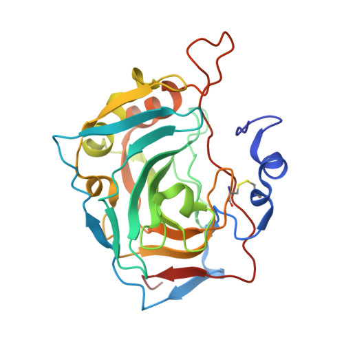
    Substituted tri- and tetrafluorobenzenesulfonamides were designed, synthesized, and evaluated as high-affinity and isoform-selective carbonic anhydrase (CA) inhibitors. Their binding affinities for recombinant human CA I, II, VA, VI, VII, XII, and XIII catalytic domains were determined by fluorescent thermal shift assay, isothermal titration calorimetry, and a stopped-flow CO2 hydration assay. Variation of the substituents at the 2-, 3-, and 4-positions yielded compounds with a broad range of binding affinities and isoform selectivities. Several 2,4-substituted-3,5,6-trifluorobenzenesulfonamides were effective CA XIII inhibitors with high selectivity over off-target CA I and CA II. 3,4-Disubstituted-2,5,6-trifluorobenzenesulfonamides bound CAs with higher affinity than 2,4-disubstituted-3,5,6-trifluorobenzenesulfonamides. Many such fluorinated benzenesulfonamides were found to be nanomolar inhibitors of CA II, CA VII, tumor-associated CA IX and CA XII, and CA XIII. X-ray crystal structures of inhibitors bound in the active sites of several CA isoforms provide structure-activity relationship information for inhibitor binding affinities and selectivity.


  • Organizational Affiliation
    • Department of Biothermodynamics and Drug Design, Institute of Biotechnology, Vilnius University, Graičiūno 8, Vilnius 02241 (Lithuania).

Macromolecule Content 

  • Total Structure Weight: 122.06 kDa 
  • Atom Count: 9,883 
  • Modeled Residue Count: 1,044 
  • Deposited Residue Count: 1,052 
  • Unique protein chains: 1

Macromolecules

Find similar proteins by:|  3D Structure
Entity ID: 1
MoleculeChains  Sequence LengthOrganismDetailsImage
Carbonic anhydrase 12
A, B, C, D
263Homo sapiensMutation(s): 0 
Gene Names: CA12
EC: 4.2.1.1
UniProt & NIH Common Fund Data Resources
Find proteins for O43570 (Homo sapiens)
Explore O43570 
Go to UniProtKB:  O43570
PHAROS:  O43570
GTEx:  ENSG00000074410 
Entity Groups
Sequence Clusters30% Identity50% Identity70% Identity90% Identity95% Identity100% Identity
UniProt GroupO43570
Sequence Annotations
Expand
Reference Sequence

Small Molecules

Ligands 3 Unique
IDChains Name / Formula / InChI Key2D Diagram3D Interactions
WWO

Query on WWO


Download:Ideal Coordinates CCD File
F [auth A],
I [auth B],
M [auth C],
P [auth D]
3-(benzylamino)-2,5,6-trifluoro-4-[(2-hydroxyethyl)sulfonyl]benzenesulfonamide
C15 H15 F3 N2 O5 S2
OLTMJLATLWYQJF-UHFFFAOYSA-N
ZN

Query on ZN


Download:Ideal Coordinates CCD File
E [auth A],
H [auth B],
L [auth C],
O [auth D]
ZINC ION
Zn
PTFCDOFLOPIGGS-UHFFFAOYSA-N
EDO

Query on EDO


Download:Ideal Coordinates CCD File
G [auth A]
J [auth B]
K [auth B]
N [auth C]
Q [auth D]
G [auth A],
J [auth B],
K [auth B],
N [auth C],
Q [auth D],
R [auth D],
S [auth D]
1,2-ETHANEDIOL
C2 H6 O2
LYCAIKOWRPUZTN-UHFFFAOYSA-N

Experimental Data & Validation

Experimental Data

  • Method: X-RAY DIFFRACTION
  • Resolution: 1.55 Å
  • R-Value Free:  0.222 (Depositor), 0.216 (DCC) 
  • R-Value Work:  0.185 (Depositor), 0.179 (DCC) 
  • R-Value Observed: 0.188 (Depositor) 
Space Group: P 1
Unit Cell:
Length ( Å )Angle ( ˚ )
a = 46.501α = 81.57
b = 66.762β = 84.21
c = 80.466γ = 86.66
Software Package:
Software NamePurpose
REFMACrefinement
MOSFLMdata reduction
SCALAdata scaling
PDB_EXTRACTdata extraction
MAR345dtbdata collection
MOLREPphasing

Structure Validation

View Full Validation Report


Ligand Structure Quality Assessment 


Entry History 

Deposition Data

Revision History  (Full details and data files)

  • Version 1.0: 2015-04-15
    Type: Initial release
  • Version 1.1: 2017-10-04
    Changes: Data collection, Refinement description
  • Version 1.2: 2023-11-08
    Changes: Data collection, Database references, Derived calculations, Refinement description
  • Version 1.3: 2024-11-13
    Changes: Structure summary