4QJ5

Structure of a fragment of human phospholipase C-beta3 delta472-581, bound to IP3 and in complex with Galphaq


Experimental Data Snapshot

  • Method: X-RAY DIFFRACTION
  • Resolution: 3.41 Å
  • R-Value Free: 0.277 
  • R-Value Work: 0.212 
  • R-Value Observed: 0.215 

wwPDB Validation   3D Report Full Report


Ligand Structure Quality Assessment 


This is version 1.4 of the entry. See complete history


Literature

Molecular mechanisms of phospholipase C beta 3 autoinhibition.

Lyon, A.M.Begley, J.A.Manett, T.D.Tesmer, J.J.

(2014) Structure 22: 1844-1854

  • DOI: https://doi.org/10.1016/j.str.2014.10.008
  • Primary Citation of Related Structures:  
    4QJ3, 4QJ4, 4QJ5

  • PubMed Abstract: 

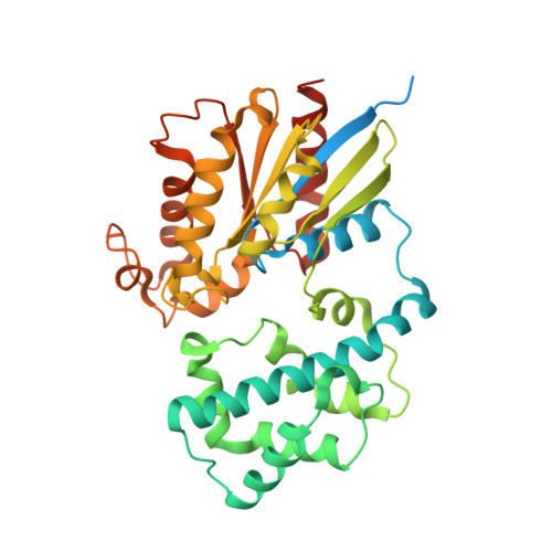
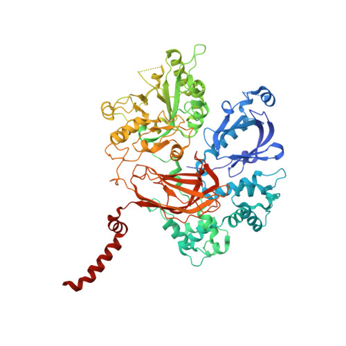
    Phospholipase C β (PLCβ) enzymes are dramatically activated by heterotrimeric G proteins. Central to this response is the robust autoinhibition of PLCβ by the X-Y linker region within its catalytic core and by the Hα2' helix in the C-terminal extension of the enzyme. The molecular mechanism of each and their mutual dependence are poorly understood. Herein, it is shown that distinct regions within the X-Y linker have specific roles in regulating activity. Most important,an acidic stretch within the linker stabilizes a lid that occludes the active site, consistent with crystal structures of variants lacking this region. Inhibition by the Hα2' helix is independent of the X-Y linker and likely regulates activity by limiting membrane interaction of the catalytic core. Full activation of PLCβ thus requires multiple independent molecular events induced by membrane association of the catalytic core and by the binding of regulatory proteins.


  • Organizational Affiliation

    Life Sciences Institute, University of Michigan, 210 Washtenaw Avenue, Ann Arbor, MI 48109-2216, USA; Department of Pharmacology, University of Michigan, 1150 W. Medical Center Drive, 1301 MSRB III, Ann Arbor, MI 48109, USA; Department of Biological Chemistry, University of Michigan, 1150 W. Medical Center Drive, RM 5301 MSRB III, Ann Arbor, MI 48109-0600, USA.


Macromolecules
Find similar proteins by:  (by identity cutoff)  |  3D Structure
Entity ID: 1
MoleculeChains Sequence LengthOrganismDetailsImage
Guanine nucleotide-binding protein G(q) subunit alpha379Mus musculusMutation(s): 0 
Gene Names: Gnaq
UniProt
Find proteins for P21279 (Mus musculus)
Explore P21279 
Go to UniProtKB:  P21279
Entity Groups  
Sequence Clusters30% Identity50% Identity70% Identity90% Identity95% Identity100% Identity
UniProt GroupP21279
Sequence Annotations
Expand
  • Reference Sequence
Find similar proteins by:  (by identity cutoff)  |  3D Structure
Entity ID: 2
MoleculeChains Sequence LengthOrganismDetailsImage
1-phosphatidylinositol 4,5-bisphosphate phosphodiesterase beta-3793Homo sapiensMutation(s): 0 
Gene Names: PLCB3
EC: 3.1.4.11
UniProt & NIH Common Fund Data Resources
Find proteins for Q01970 (Homo sapiens)
Explore Q01970 
Go to UniProtKB:  Q01970
PHAROS:  Q01970
GTEx:  ENSG00000149782 
Entity Groups  
Sequence Clusters30% Identity50% Identity70% Identity90% Identity95% Identity100% Identity
UniProt GroupQ01970
Sequence Annotations
Expand
  • Reference Sequence
Small Molecules
Ligands 5 Unique
IDChains Name / Formula / InChI Key2D Diagram3D Interactions
GDP
Query on GDP

Download Ideal Coordinates CCD File 
C [auth A]GUANOSINE-5'-DIPHOSPHATE
C10 H15 N5 O11 P2
QGWNDRXFNXRZMB-UUOKFMHZSA-N
I3P
Query on I3P

Download Ideal Coordinates CCD File 
G [auth B]D-MYO-INOSITOL-1,4,5-TRIPHOSPHATE
C6 H15 O15 P3
MMWCIQZXVOZEGG-XJTPDSDZSA-N
ALF
Query on ALF

Download Ideal Coordinates CCD File 
D [auth A]TETRAFLUOROALUMINATE ION
Al F4
UYOMQIYKOOHAMK-UHFFFAOYSA-J
CA
Query on CA

Download Ideal Coordinates CCD File 
F [auth B]CALCIUM ION
Ca
BHPQYMZQTOCNFJ-UHFFFAOYSA-N
MG
Query on MG

Download Ideal Coordinates CCD File 
E [auth A]MAGNESIUM ION
Mg
JLVVSXFLKOJNIY-UHFFFAOYSA-N
Experimental Data & Validation

Experimental Data

  • Method: X-RAY DIFFRACTION
  • Resolution: 3.41 Å
  • R-Value Free: 0.277 
  • R-Value Work: 0.212 
  • R-Value Observed: 0.215 
  • Space Group: C 1 2 1
Unit Cell:
Length ( Å )Angle ( ˚ )
a = 205.918α = 90
b = 89.869β = 101.8
c = 93.261γ = 90
Software Package:
Software NamePurpose
HKL-2000data collection
REFMACrefinement
DENZOdata reduction
HKL-2000data scaling
REFMACphasing

Structure Validation

View Full Validation Report


Ligand Structure Quality Assessment 


Entry History 

Deposition Data

Revision History  (Full details and data files)

  • Version 1.0: 2014-10-22
    Type: Initial release
  • Version 1.1: 2014-12-24
    Changes: Database references
  • Version 1.2: 2015-02-11
    Changes: Database references
  • Version 1.3: 2017-08-02
    Changes: Data collection, Refinement description, Source and taxonomy
  • Version 1.4: 2023-09-20
    Changes: Data collection, Database references, Derived calculations, Refinement description