Inactivation of the Mycobacterium tuberculosis Antigen 85 Complex by Covalent, Allosteric Inhibitors.
Favrot, L., Lajiness, D.H., Ronning, D.R.(2014) J Biological Chem 289: 25031-25040
- PubMed: 25028518 Search on PubMedSearch on PubMed Central
- DOI: https://doi.org/10.1074/jbc.M114.582445
- Primary Citation Related Structures:
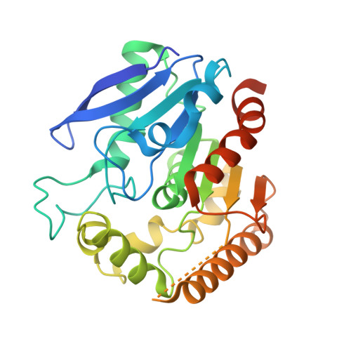
4QDO, 4QDT, 4QDU, 4QDX, 4QDZ, 4QE3, 4QEK - PubMed Abstract:
The rise of multidrug-resistant and totally drug-resistant tuberculosis and the association with an increasing number of HIV-positive patients developing tuberculosis emphasize the necessity to find new antitubercular targets and drugs. The antigen 85 (Ag85) complex from Mycobacterium tuberculosis plays important roles in the biosynthesis of major components of the mycobacterial cell envelope. For this reason, Ag85 has emerged as an attractive drug target. Recently, ebselen was identified as an effective inhibitor of the Ag85 complex through covalent modification of a cysteine residue proximal to the Ag85 active site and is therefore a covalent, allosteric inhibitor. To expand the understanding of this process, we have solved the x-ray crystal structures of Ag85C covalently modified with ebselen and other thiol-reactive compounds, p-chloromercuribenzoic acid and iodoacetamide, as well as the structure of a cysteine to glycine mutant. All four structures confirm that chemical modification or mutation at this particular cysteine residue leads to the disruption of the active site hydrogen-bonded network essential for Ag85 catalysis. We also describe x-ray crystal structures of Ag85C single mutants within the catalytic triad and show that a mutation of any one of these three residues promotes the same conformational change observed in the cysteine-modified forms. These results provide evidence for active site dynamics that may afford new strategies for the development of selective and potent Ag85 inhibitors.
- From the Department of Chemistry and Biochemistry, University of Toledo, Toledo, Ohio 43606-3390.
Organizational Affiliation: