


Dcps in complex with covalent ligands targeting Tyrosines
Liu, S.To be published.
Experimental Data Snapshot
Entity ID: 1 | |||||
|---|---|---|---|---|---|
| Molecule | Chains | Sequence Length | Organism | Details | Image |
| m7GpppX diphosphatase | 337 | Homo sapiens | Mutation(s): 0 Gene Names: DCPS, DCS1, HINT5, HSPC015 EC: 3.6.1.59 | 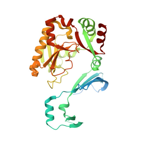 | |
UniProt & NIH Common Fund Data Resources | |||||
PHAROS: Q96C86 GTEx: ENSG00000110063 | |||||
Entity Groups | |||||
| Sequence Clusters | 30% Identity50% Identity70% Identity90% Identity95% Identity100% Identity | ||||
| UniProt Group | Q96C86 | ||||
Sequence AnnotationsExpand | |||||
Reference Sequence | |||||
| Ligands 3 Unique | |||||
|---|---|---|---|---|---|
| ID | Chains | Name / Formula / InChI Key | 2D Diagram | 3D Interactions | |
| 30U Download:Ideal Coordinates CCD File | E [auth A], N [auth D] | 4-{[(2,4-diaminoquinazolin-5-yl)oxy]methyl}benzenesulfonic acid C15 H14 N4 O4 S AVKNBINNLVQZGS-UHFFFAOYSA-N |  | ||
| PO4 Download:Ideal Coordinates CCD File | F [auth A] G [auth A] H [auth A] I [auth B] J [auth B] | PHOSPHATE ION O4 P NBIIXXVUZAFLBC-UHFFFAOYSA-K |  | ||
| GOL Download:Ideal Coordinates CCD File | Q [auth D] | GLYCEROL C3 H8 O3 PEDCQBHIVMGVHV-UHFFFAOYSA-N |  | ||
| Length ( Å ) | Angle ( ˚ ) |
|---|---|
| a = 101.09 | α = 90 |
| b = 105.51 | β = 90 |
| c = 140 | γ = 90 |
| Software Name | Purpose |
|---|---|
| JDirector | data collection |
| BUSTER | refinement |
| autoPROC | data scaling |
| Aimless | data scaling |
| BUSTER | phasing |


