4QCC

Structure of a cube-shaped, highly porous protein cage designed by fusing symmetric oligomeric domains


Experimental Data Snapshot

  • Method: X-RAY DIFFRACTION
  • Resolution: 7.08 Å
  • R-Value Free: 0.317 
  • R-Value Work: 0.283 
  • R-Value Observed: 0.284 

wwPDB Validation   3D Report Full Report


This is version 1.3 of the entry. See complete history


Literature

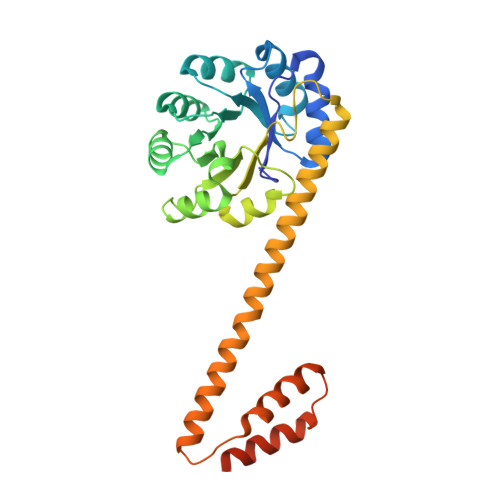
Structure of a designed protein cage that self-assembles into a highly porous cube.

Lai, Y.T.Reading, E.Hura, G.L.Tsai, K.L.Laganowsky, A.Asturias, F.J.Tainer, J.A.Robinson, C.V.Yeates, T.O.

(2014) Nat Chem 6: 1065-1071

  • DOI: https://doi.org/10.1038/nchem.2107
  • Primary Citation of Related Structures:  
    4QCC

  • PubMed Abstract: 

    Natural proteins can be versatile building blocks for multimeric, self-assembling structures. Yet, creating protein-based assemblies with specific geometries and chemical properties remains challenging. Highly porous materials represent particularly interesting targets for designed assembly. Here, we utilize a strategy of fusing two natural protein oligomers using a continuous alpha-helical linker to design a novel protein that self assembles into a 750 kDa, 225 Å diameter, cube-shaped cage with large openings into a 130 Å diameter inner cavity. A crystal structure of the cage showed atomic-level agreement with the designed model, while electron microscopy, native mass spectrometry and small angle X-ray scattering revealed alternative assembly forms in solution. These studies show that accurate design of large porous assemblies with specific shapes is feasible, while further specificity improvements will probably require limiting flexibility to select against alternative forms. These results provide a foundation for the design of advanced materials with applications in bionanotechnology, nanomedicine and material sciences.


  • Organizational Affiliation

    UCLA-DOE Institute for Genomics and Proteomics, University of California, Los Angeles, California 90095, USA.


Macromolecules
Find similar proteins by:  (by identity cutoff)  |  3D Structure
Entity ID: 1
MoleculeChains Sequence LengthOrganismDetailsImage
2-dehydro-3-deoxy-6-phosphogalactonate aldolase, peptidyl-prolyl cis-trans isomerase chimera
A, B
291Escherichia coliMutation(s): 0 
Gene Names: dgoAfkpA
EC: 4.1.2.21
UniProt
Find proteins for P45523 (Escherichia coli (strain K12))
Explore P45523 
Go to UniProtKB:  P45523
Find proteins for Q6BF16 (Escherichia coli (strain K12))
Explore Q6BF16 
Go to UniProtKB:  Q6BF16
Entity Groups  
Sequence Clusters30% Identity50% Identity70% Identity90% Identity95% Identity100% Identity
UniProt GroupsQ6BF16P45523
Sequence Annotations
Expand
  • Reference Sequence
Experimental Data & Validation

Experimental Data

  • Method: X-RAY DIFFRACTION
  • Resolution: 7.08 Å
  • R-Value Free: 0.317 
  • R-Value Work: 0.283 
  • R-Value Observed: 0.284 
  • Space Group: F 2 3
Unit Cell:
Length ( Å )Angle ( ˚ )
a = 272.68α = 90
b = 272.68β = 90
c = 272.68γ = 90
Software Package:
Software NamePurpose
XSCALEdata scaling
PHASERphasing
REFMACrefinement
PDB_EXTRACTdata extraction

Structure Validation

View Full Validation Report


Entry History 

Deposition Data

Revision History  (Full details and data files)

  • Version 1.0: 2014-11-19
    Type: Initial release
  • Version 1.1: 2014-12-03
    Changes: Database references
  • Version 1.2: 2017-07-26
    Changes: Refinement description, Source and taxonomy
  • Version 1.3: 2024-02-28
    Changes: Data collection, Database references, Refinement description