4Q7Y | pdb_00004q7y

Neutrophil serine protease 4 (PRSS57) apo form 2


Experimental Data Snapshot

  • Method: X-RAY DIFFRACTION
  • Resolution: 2.70 Å
  • R-Value Free: 
    0.248 (Depositor), 0.244 (DCC) 
  • R-Value Work: 
    0.187 (Depositor), 0.186 (DCC) 
  • R-Value Observed: 
    0.193 (Depositor) 

Starting Model: experimental
View more details

wwPDB Validation 3D Report Full Report

Validation slider image for 4Q7Y

Ligand Structure Quality Assessment 


This is version 2.2 of the entry. See complete history

Literature

Structures of neutrophil serine protease 4 reveal an unusual mechanism of substrate recognition by a trypsin-fold protease.

Lin, S.J.Dong, K.C.Eigenbrot, C.van Lookeren Campagne, M.Kirchhofer, D.

(2014) Structure 22: 1333-1340

  • DOI: https://doi.org/10.1016/j.str.2014.07.008
  • Primary Citation Related Structures: 
    4Q7X, 4Q7Y, 4Q7Z, 4Q80

  • PubMed Abstract: 

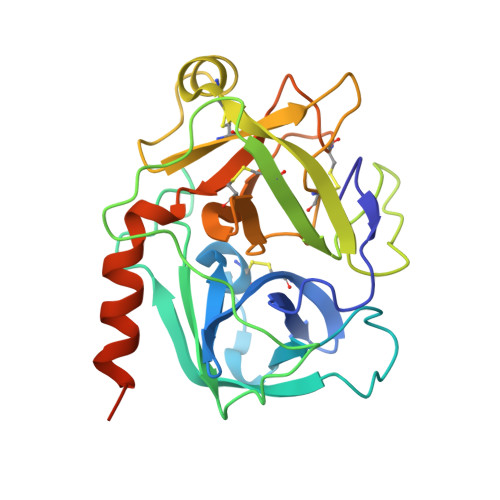
    Trypsin-fold proteases, the largest mammalian protease family, are classified by their primary substrate specificity into one of three categories, trypsin-like, chymotrypsin-like, and elastase-like, based on key structural features of their active site. However, the recently discovered neutrophil serine protease 4 (NSP4, also known as PRSS57) presents a paradox: NSP4 exhibits a trypsin-like specificity for cleaving substrates after arginine residues, but it bears elastase-like specificity determining residues in the active site. Here we show that NSP4 has a fully occluded S1 pocket and that the substrate P1-arginine adopts a noncanonical "up" conformation stabilized by a solvent-exposed H-bond network. This uncommon arrangement, conserved in all NSP4 orthologs, enables NSP4 to process substrates after both arginine as well as post-translationally modified arginine residues, such as methylarginine and citrulline. These findings establish a distinct paradigm for substrate recognition by a trypsin-fold protease and provide insights into the function of NSP4.


  • Organizational Affiliation
    • Department of Early Discovery Biochemistry, Genentech, Inc., 1 DNA Way, South San Francisco, CA 94080, USA.

Macromolecule Content 

  • Total Structure Weight: 27.63 kDa 
  • Atom Count: 1,850 
  • Modeled Residue Count: 234 
  • Deposited Residue Count: 250 
  • Unique protein chains: 1

Macromolecules

Find similar proteins by:|  3D Structure
Entity ID: 1
MoleculeChains  Sequence LengthOrganismDetailsImage
Serine protease 57250Homo sapiensMutation(s): 0 
Gene Names: PRSS57PRSSL1UNQ782/PRO1599
EC: 3.4.21
UniProt & NIH Common Fund Data Resources
Find proteins for Q6UWY2 (Homo sapiens)
Explore Q6UWY2 
Go to UniProtKB:  Q6UWY2
PHAROS:  Q6UWY2
GTEx:  ENSG00000185198 
Entity Groups
Sequence Clusters30% Identity50% Identity70% Identity90% Identity95% Identity100% Identity
UniProt GroupQ6UWY2
Glycosylation
Glycosylation Sites: 2Go to GlyGen: Q6UWY2-1
Sequence Annotations
Expand
Reference Sequence

Oligosaccharides

Help  
Entity ID: 2
MoleculeChains Length2D Diagram GlycosylationD Interactions
alpha-L-fucopyranose-(1-6)-2-acetamido-2-deoxy-beta-D-glucopyranose
B
2N-Glycosylation
Glycosylation Resources
GlyTouCan: G86851RC
GlyCosmos: G86851RC
GlyGen: G86851RC

Small Molecules

Experimental Data & Validation

Experimental Data

  • Method: X-RAY DIFFRACTION
  • Resolution: 2.70 Å
  • R-Value Free:  0.248 (Depositor), 0.244 (DCC) 
  • R-Value Work:  0.187 (Depositor), 0.186 (DCC) 
  • R-Value Observed: 0.193 (Depositor) 
Space Group: P 63
Unit Cell:
Length ( Å )Angle ( ˚ )
a = 86.636α = 90
b = 86.636β = 90
c = 69.351γ = 120
Software Package:
Software NamePurpose
BOSdata collection
BUSTERrefinement
HKL-2000data reduction
HKL-2000data scaling

Structure Validation

View Full Validation Report


Ligand Structure Quality Assessment 


Entry History 

Deposition Data

Revision History  (Full details and data files)

  • Version 1.0: 2014-09-03
    Type: Initial release
  • Version 1.1: 2014-09-17
    Changes: Database references
  • Version 1.2: 2017-11-22
    Changes: Refinement description
  • Version 2.0: 2020-07-29
    Type: Remediation
    Reason: Carbohydrate remediation
    Changes: Atomic model, Data collection, Derived calculations, Structure summary
  • Version 2.1: 2023-09-20
    Changes: Data collection, Database references, Refinement description, Structure summary
  • Version 2.2: 2024-11-20
    Changes: Structure summary