4Q7O

The crystal structure of an immunity protein NMB0503 from Neisseria meningitidis MC58


Experimental Data Snapshot

  • Method: X-RAY DIFFRACTION
  • Resolution: 1.45 Å
  • R-Value Free: 0.189 
  • R-Value Work: 0.149 
  • R-Value Observed: 0.151 

wwPDB Validation   3D Report Full Report


This is version 1.2 of the entry. See complete history


Literature

The structure of a contact-dependent growth-inhibition (CDI) immunity protein from Neisseria meningitidis MC58.

Tan, K.Johnson, P.M.Stols, L.Boubion, B.Eschenfeldt, W.Babnigg, G.Hayes, C.S.Joachimiak, A.Goulding, C.W.

(2015) Acta Crystallogr F Struct Biol Commun 71: 702-709

  • DOI: https://doi.org/10.1107/S2053230X15006585
  • Primary Citation of Related Structures:  
    4Q7O

  • PubMed Abstract: 

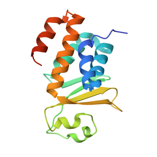
    Contact-dependent growth inhibition (CDI) is an important mechanism of intercellular competition between neighboring Gram-negative bacteria. CDI systems encode large surface-exposed CdiA effector proteins that carry a variety of C-terminal toxin domains (CdiA-CTs). All CDI(+) bacteria also produce CdiI immunity proteins that specifically bind to the cognate CdiA-CT and neutralize its toxin activity to prevent auto-inhibition. Here, the X-ray crystal structure of a CdiI immunity protein from Neisseria meningitidis MC58 is presented at 1.45 Å resolution. The CdiI protein has structural homology to the Whirly family of RNA-binding proteins, but appears to lack the characteristic nucleic acid-binding motif of this family. Sequence homology suggests that the cognate CdiA-CT is related to the eukaryotic EndoU family of RNA-processing enzymes. A homology model is presented of the CdiA-CT based on the structure of the XendoU nuclease from Xenopus laevis. Molecular-docking simulations predict that the CdiA-CT toxin active site is occluded upon binding to the CdiI immunity protein. Together, these observations suggest that the immunity protein neutralizes toxin activity by preventing access to RNA substrates.


  • Organizational Affiliation

    Midwest Center for Structural Genomics, Argonne National Laboratory, Argonne, IL 60439, USA.


Macromolecules
Find similar proteins by:  (by identity cutoff)  |  3D Structure
Entity ID: 1
MoleculeChains Sequence LengthOrganismDetailsImage
Immunity protein
A, B
146Neisseria meningitidis MC58Mutation(s): 0 
Gene Names: NMB0503
UniProt
Find proteins for Q7DDN8 (Neisseria meningitidis serogroup B (strain MC58))
Explore Q7DDN8 
Go to UniProtKB:  Q7DDN8
Entity Groups  
Sequence Clusters30% Identity50% Identity70% Identity90% Identity95% Identity100% Identity
UniProt GroupQ7DDN8
Sequence Annotations
Expand
  • Reference Sequence
Small Molecules
Ligands 2 Unique
IDChains Name / Formula / InChI Key2D Diagram3D Interactions
BR
Query on BR

Download Ideal Coordinates CCD File 
C [auth A]
D [auth A]
E [auth A]
F [auth A]
G [auth A]
C [auth A],
D [auth A],
E [auth A],
F [auth A],
G [auth A],
H [auth A],
K [auth B],
L [auth B],
M [auth B],
N [auth B],
O [auth B],
P [auth B]
BROMIDE ION
Br
CPELXLSAUQHCOX-UHFFFAOYSA-M
FMT
Query on FMT

Download Ideal Coordinates CCD File 
I [auth A],
J [auth A],
Q [auth B]
FORMIC ACID
C H2 O2
BDAGIHXWWSANSR-UHFFFAOYSA-N
Experimental Data & Validation

Experimental Data

  • Method: X-RAY DIFFRACTION
  • Resolution: 1.45 Å
  • R-Value Free: 0.189 
  • R-Value Work: 0.149 
  • R-Value Observed: 0.151 
  • Space Group: P 1 21 1
Unit Cell:
Length ( Å )Angle ( ˚ )
a = 45.383α = 90
b = 53.532β = 98.04
c = 59.699γ = 90
Software Package:
Software NamePurpose
SBC-Collectdata collection
SHELXDphasing
MLPHAREphasing
DMmodel building
ARPmodel building
WARPmodel building
HKL-3000phasing
PHENIXrefinement
HKL-3000data reduction
HKL-3000data scaling
DMphasing

Structure Validation

View Full Validation Report


Entry History 

Revision History  (Full details and data files)

  • Version 1.0: 2014-05-14
    Type: Initial release
  • Version 1.1: 2015-07-01
    Changes: Database references
  • Version 1.2: 2024-02-28
    Changes: Data collection, Database references, Derived calculations