


Crystal structure of plectin 1a actin-binding domain
Song, J.-G., Kostan, J., Grishkovskaya, I., Djinovic-Carugo, K.To be published.
Experimental Data Snapshot
wwPDB Validation 3D Report Full Report

Entity ID: 1 | |||||
|---|---|---|---|---|---|
| Molecule | Chains | Sequence Length | Organism | Details | Image |
| Plectin | 226 | Homo sapiens | Mutation(s): 0 | 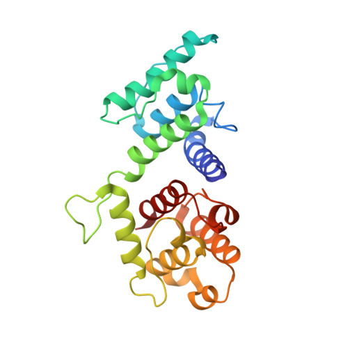 | |
UniProt & NIH Common Fund Data Resources | |||||
PHAROS: Q15149 GTEx: ENSG00000178209 | |||||
Entity Groups | |||||
| Sequence Clusters | 30% Identity50% Identity70% Identity90% Identity95% Identity100% Identity | ||||
| UniProt Group | Q15149 | ||||
Sequence AnnotationsExpand | |||||
Reference Sequence | |||||
| Length ( Å ) | Angle ( ˚ ) |
|---|---|
| a = 41.85 | α = 90 |
| b = 159.823 | β = 90 |
| c = 184.27 | γ = 90 |
| Software Name | Purpose |
|---|---|
| PHENIX | refinement |


