4Q3A | pdb_00004q3a

PylD cocrystallized with L-Lysine-Ne-3S-methyl-L-ornithine and NAD+


Experimental Data Snapshot

  • Method: X-RAY DIFFRACTION
  • Resolution: 2.20 Å
  • R-Value Free: 
    0.204 (Depositor), 0.210 (DCC) 
  • R-Value Work: 
    0.169 (Depositor), 0.178 (DCC) 
  • R-Value Observed: 
    0.171 (Depositor) 

Starting Model: experimental
View more details

wwPDB Validation 3D Report Full Report

Validation slider image for 4Q3A

Ligand Structure Quality Assessment 


This is version 1.4 of the entry. See complete history

Literature

The Formation of Pyrroline and Tetrahydropyridine Rings in Amino Acids Catalyzed by Pyrrolysine Synthase (PylD).

Quitterer, F.Beck, P.Bacher, A.Groll, M.

(2014) Angew Chem Int Ed Engl 53: 8150-8153

  • DOI: https://doi.org/10.1002/anie.201402595
  • Primary Citation Related Structures: 
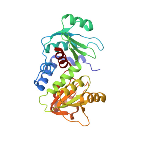
    4Q39, 4Q3A, 4Q3B, 4Q3C, 4Q3D, 4Q3E

  • PubMed Abstract: 

    The dehydrogenase PylD catalyzes the ultimate step of the pyrrolysine pathway by converting the isopeptide L-lysine-Nε-3R-methyl-D-ornithine to the 22nd proteinogenic amino acid. In this study, we demonstrate how PylD can be harnessed to oxidize various isopeptides to novel amino acids by combining chemical synthesis with enzyme kinetics and X-ray crystallography. The data enable a detailed description of the PylD reaction trajectory for the biosynthesis of pyrroline and tetrahydropyridine rings as constituents of pyrrolysine analogues.


  • Organizational Affiliation
    • Center for Integrated Protein Science Munich (CIPSM), Lehrstuhl für Biochemie, Technische Universität München, Lichtenbergstrasse 4, 85748 Garching (Germany).

Macromolecule Content 

  • Total Structure Weight: 118.33 kDa 
  • Atom Count: 8,575 
  • Modeled Residue Count: 1,037 
  • Deposited Residue Count: 1,040 
  • Unique protein chains: 1

Macromolecules

Find similar proteins by:|  3D Structure
Entity ID: 1
MoleculeChains  Sequence LengthOrganismDetailsImage
PYLD, pyrrolysine synthase
A, B, C, D
260Methanosarcina barkeri str. FusaroMutation(s): 0 
Gene Names: Mbar_A0835
EC: 1.4.1
UniProt
Find proteins for Q46E80 (Methanosarcina barkeri (strain Fusaro / DSM 804))
Explore Q46E80 
Go to UniProtKB:  Q46E80
Entity Groups
Sequence Clusters30% Identity50% Identity70% Identity90% Identity95% Identity100% Identity
UniProt GroupQ46E80
Sequence Annotations
Expand
Reference Sequence

Small Molecules

Ligands 8 Unique
IDChains Name / Formula / InChI Key2D Diagram3D Interactions
NAI

Query on NAI


Download:Ideal Coordinates CCD File
E [auth A],
OA [auth C],
T [auth B],
WA [auth D]
1,4-DIHYDRONICOTINAMIDE ADENINE DINUCLEOTIDE
C21 H29 N7 O14 P2
BOPGDPNILDQYTO-NNYOXOHSSA-N
2YC

Query on 2YC


Download:Ideal Coordinates CCD File
H [auth A],
RA [auth C],
W [auth B],
ZA [auth D]
N~6~-{[(2S,3S)-3-methyl-3,4-dihydro-2H-pyrrol-2-yl]carbonyl}-L-lysine
C12 H21 N3 O3
ZFOMKMMPBOQKMC-GUBZILKMSA-N
PEG

Query on PEG


Download:Ideal Coordinates CCD File
FA [auth B],
GA [auth B],
P [auth A]
DI(HYDROXYETHYL)ETHER
C4 H10 O3
MTHSVFCYNBDYFN-UHFFFAOYSA-N
SO4

Query on SO4


Download:Ideal Coordinates CCD File
DA [auth B],
EA [auth B]
SULFATE ION
O4 S
QAOWNCQODCNURD-UHFFFAOYSA-L
GOL

Query on GOL


Download:Ideal Coordinates CCD File
AA [auth B]
BA [auth B]
CA [auth B]
I [auth A]
J [auth A]
AA [auth B],
BA [auth B],
CA [auth B],
I [auth A],
J [auth A],
K [auth A],
L [auth A],
M [auth A],
N [auth A],
O [auth A],
SA [auth C],
X [auth B],
Y [auth B],
Z [auth B]
GLYCEROL
C3 H8 O3
PEDCQBHIVMGVHV-UHFFFAOYSA-N
EDO

Query on EDO


Download:Ideal Coordinates CCD File
HA [auth B]
IA [auth B]
JA [auth B]
KA [auth B]
LA [auth B]
HA [auth B],
IA [auth B],
JA [auth B],
KA [auth B],
LA [auth B],
MA [auth B],
NA [auth B],
Q [auth A],
R [auth A],
S [auth A],
TA [auth C],
UA [auth C],
VA [auth C]
1,2-ETHANEDIOL
C2 H6 O2
LYCAIKOWRPUZTN-UHFFFAOYSA-N
MG

Query on MG


Download:Ideal Coordinates CCD File
G [auth A],
QA [auth C],
V [auth B],
YA [auth D]
MAGNESIUM ION
Mg
JLVVSXFLKOJNIY-UHFFFAOYSA-N
NA

Query on NA


Download:Ideal Coordinates CCD File
F [auth A],
PA [auth C],
U [auth B],
XA [auth D]
SODIUM ION
Na
FKNQFGJONOIPTF-UHFFFAOYSA-N

Experimental Data & Validation

Experimental Data

  • Method: X-RAY DIFFRACTION
  • Resolution: 2.20 Å
  • R-Value Free:  0.204 (Depositor), 0.210 (DCC) 
  • R-Value Work:  0.169 (Depositor), 0.178 (DCC) 
  • R-Value Observed: 0.171 (Depositor) 
Space Group: P 21 21 2
Unit Cell:
Length ( Å )Angle ( ˚ )
a = 87.49α = 90
b = 259.85β = 90
c = 48.66γ = 90
Software Package:
Software NamePurpose
XDSdata scaling
PHASERphasing
REFMACrefinement
XDSdata reduction
XSCALEdata scaling

Structure Validation

View Full Validation Report


Ligand Structure Quality Assessment 


Entry History 

Deposition Data

Revision History  (Full details and data files)

  • Version 1.0: 2014-04-23
    Type: Initial release
  • Version 1.1: 2014-07-02
    Changes: Database references
  • Version 1.2: 2014-08-20
    Changes: Database references
  • Version 1.3: 2023-09-20
    Changes: Data collection, Database references, Derived calculations, Refinement description
  • Version 1.4: 2024-10-16
    Changes: Structure summary