4Q24 | pdb_00004q24

Crystal structure of Cyclo(L-leucyl-L-phenylalanyl) synthase


Experimental Data Snapshot

  • Method: X-RAY DIFFRACTION
  • Resolution: 2.90 Å
  • R-Value Free: 
    0.253 (Depositor), 0.224 (DCC) 
  • R-Value Work: 
    0.201 (Depositor) 
  • R-Value Observed: 
    0.203 (Depositor) 

Starting Model: experimental
View more details

wwPDB Validation 3D Report Full Report

Validation slider image for 4Q24

Ligand Structure Quality Assessment 


This is version 1.4 of the entry. See complete history

Literature

Unravelling the mechanism of non-ribosomal peptide synthesis by cyclodipeptide synthases.

Moutiez, M.Schmitt, E.Seguin, J.Thai, R.Favry, E.Belin, P.Mechulam, Y.Gondry, M.

(2014) Nat Commun 5: 5141-5141

  • DOI: https://doi.org/10.1038/ncomms6141
  • Primary Citation Related Structures: 
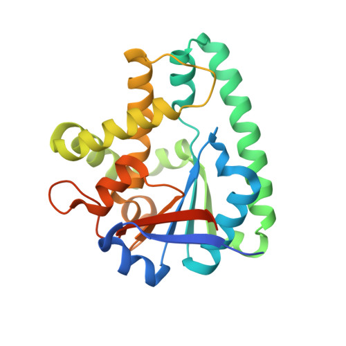
    4Q24

  • PubMed Abstract: 

    Cyclodipeptide synthases form cyclodipeptides from two aminoacyl transfer RNAs. They use a ping-pong mechanism that begins with transfer of the aminoacyl moiety of the first aminoacyl tRNA onto a conserved serine, yielding an aminoacyl enzyme. Combining X-ray crystallography, site-directed mutagenesis and affinity labelling of the cyclodipeptide synthase AlbC, we demonstrate that the covalent intermediate reacts with the aminoacyl moiety of the second aminoacyl tRNA, forming a dipeptidyl enzyme, and identify the aminoacyl-binding sites of the aminoacyl tRNAs.


  • Organizational Affiliation
    • Service d'Ingénierie Moléculaire des Protéines, iBiTec-S, Commissariat à l'Energie Atomique et aux Energies Alternatives (CEA), 91191 Gif-sur-Yvette, France.

Macromolecule Content 

  • Total Structure Weight: 28.23 kDa 
  • Atom Count: 1,804 
  • Modeled Residue Count: 224 
  • Deposited Residue Count: 247 
  • Unique protein chains: 1

Macromolecules

Find similar proteins by:|  3D Structure
Entity ID: 1
MoleculeChains  Sequence LengthOrganismDetailsImage
Cyclo(L-leucyl-L-phenylalanyl) synthase247Streptomyces nourseiMutation(s): 1 
Gene Names: albC
EC: 2.3.2.20
UniProt
Find proteins for Q8GED7 (Streptomyces noursei)
Explore Q8GED7 
Go to UniProtKB:  Q8GED7
Entity Groups
Sequence Clusters30% Identity50% Identity70% Identity90% Identity95% Identity100% Identity
UniProt GroupQ8GED7
Sequence Annotations
Expand
Reference Sequence

Small Molecules

Ligands 1 Unique
IDChains Name / Formula / InChI Key2D Diagram3D Interactions
XVE

Query on XVE


Download:Ideal Coordinates CCD File
B [auth A]PHENYLMETHYL N-[(2S)-4-CHLORO-3-OXO-1-PHENYL-BUTAN-2-YL]CARBAMATE
C18 H18 Cl N O3
OYHLRJGDELITAF-INIZCTEOSA-N

Experimental Data & Validation

Experimental Data

  • Method: X-RAY DIFFRACTION
  • Resolution: 2.90 Å
  • R-Value Free:  0.253 (Depositor), 0.224 (DCC) 
  • R-Value Work:  0.201 (Depositor) 
  • R-Value Observed: 0.203 (Depositor) 
Space Group: P 32 2 1
Unit Cell:
Length ( Å )Angle ( ˚ )
a = 86.968α = 90
b = 86.968β = 90
c = 70.738γ = 120
Software Package:
Software NamePurpose
PHASERphasing
BUSTERrefinement
XDSdata reduction
XSCALEdata scaling

Structure Validation

View Full Validation Report


Ligand Structure Quality Assessment 


Entry History 

Deposition Data

Revision History  (Full details and data files)

  • Version 1.0: 2014-10-08
    Type: Initial release
  • Version 1.1: 2014-12-31
    Changes: Database references
  • Version 1.2: 2017-11-22
    Changes: Refinement description
  • Version 1.3: 2023-09-20
    Changes: Data collection, Database references, Derived calculations, Refinement description
  • Version 1.4: 2024-12-25
    Changes: Advisory, Derived calculations, Structure summary