Structure and mechanism of a dehydratase/decarboxylase enzyme couple involved in polyketide beta-methyl branch incorporation.
Nair, A.V., Robson, A., Ackrill, T.D., Till, M., Byrne, M.J., Back, C.R., Tiwari, K., Davies, J.A., Willis, C.L., Race, P.R.(2020) Sci Rep 10: 15323-15323
- PubMed: 32948786 Search on PubMedSearch on PubMed Central
- DOI: https://doi.org/10.1038/s41598-020-71850-w
- Primary Citation Related Structures:
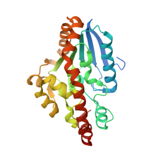
4Q1G, 4Q1H, 4Q1I, 4Q1J, 4Q1K - PubMed Abstract:
Complex polyketides of bacterial origin are biosynthesised by giant assembly-line like megaenzymes of the type 1 modular polyketide synthase (PKS) class. The trans-AT family of modular PKSs, whose biosynthetic frameworks diverge significantly from those of the archetypal cis-AT type systems represent a new paradigm in natural product enzymology. One of the most distinctive enzymatic features common to trans-AT PKSs is their ability to introduce methyl groups at positions β to the thiol ester in the growing polyketide chain. This activity is achieved through the action of a five protein HCS cassette, comprising a ketosynthase, a 3-hydroxy-3-methylglutaryl-CoA synthase, a dehydratase, a decarboxylase and a dedicated acyl carrier protein. Here we report a molecular level description, achieved using a combination of X-ray crystallography, in vitro enzyme assays and site-directed mutagenesis, of the bacillaene synthase dehydratase/decarboxylase enzyme couple PksH/PksI, responsible for the final two steps in β-methyl branch installation in this trans-AT PKS. Our work provides detailed mechanistic insight into this biosynthetic peculiarity and establishes a molecular framework for HCS cassette enzyme exploitation and manipulation, which has future potential value in guiding efforts in the targeted synthesis of functionally optimised 'non-natural' natural products.
- School of Biochemistry, University of Bristol, University Walk, Bristol, BS8 1TD, UK.
Organizational Affiliation: