


The crystal structure of a solute binding protein from Bacillus anthracis str. Ames in complex with quorum-sensing signal autoinducer-2 (AI-2).
Tan, K., Gu, M., Kwon, K., Anderson, W.F., Joachimiak, A.To be published.
Experimental Data Snapshot
Entity ID: 1 | |||||
|---|---|---|---|---|---|
| Molecule | Chains | Sequence Length | Organism | Details | Image |
| sugar ABC transporter, sugar-binding protein | 324 | Bacillus anthracis str. Ames | Mutation(s): 0 Gene Names: BA_2975, BAS2763, GBAA_2975 | 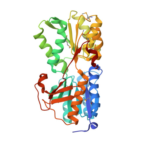 | |
UniProt | |||||
Entity Groups | |||||
| Sequence Clusters | 30% Identity50% Identity70% Identity90% Identity95% Identity100% Identity | ||||
| UniProt Group | A0A6H3AKG3 | ||||
Sequence AnnotationsExpand | |||||
Reference Sequence | |||||
| Ligands 3 Unique | |||||
|---|---|---|---|---|---|
| ID | Chains | Name / Formula / InChI Key | 2D Diagram | 3D Interactions | |
| PAV Download:Ideal Coordinates CCD File | B [auth A] | (2R,4S)-2-methyl-2,3,3,4-tetrahydroxytetrahydrofuran C5 H10 O5 BVIYGXUQVXBHQS-IUYQGCFVSA-N |  | ||
| EDO Download:Ideal Coordinates CCD File | C [auth A] D [auth A] E [auth A] F [auth A] G [auth A] | 1,2-ETHANEDIOL C2 H6 O2 LYCAIKOWRPUZTN-UHFFFAOYSA-N |  | ||
| CL Download:Ideal Coordinates CCD File | J [auth A] | CHLORIDE ION Cl VEXZGXHMUGYJMC-UHFFFAOYSA-M |  | ||
| Modified Residues 1 Unique | |||||
|---|---|---|---|---|---|
| ID | Chains | Type | Formula | 2D Diagram | Parent |
| MSE Query on MSE | A | L-PEPTIDE LINKING | C5 H11 N O2 Se |  | MET |
| Length ( Å ) | Angle ( ˚ ) |
|---|---|
| a = 39.005 | α = 90 |
| b = 58.067 | β = 95.58 |
| c = 63.194 | γ = 90 |
| Software Name | Purpose |
|---|---|
| SBC-Collect | data collection |
| SHELXD | phasing |
| MLPHARE | phasing |
| DM | model building |
| ARP | model building |
| WARP | model building |
| HKL-3000 | phasing |
| PHENIX | refinement |
| HKL-3000 | data reduction |
| HKL-3000 | data scaling |
| DM | phasing |


