4PYG | pdb_00004pyg

Transglutaminase2 complexed with GTP


Experimental Data Snapshot

  • Method: X-RAY DIFFRACTION
  • Resolution: 2.80 Å
  • R-Value Free: 
    0.319 (Depositor), 0.309 (DCC) 
  • R-Value Work: 
    0.236 (Depositor), 0.230 (DCC) 
  • R-Value Observed: 
    0.240 (Depositor) 

wwPDB Validation 3D Report Full Report

Validation slider image for 4PYG

Ligand Structure Quality Assessment 


This is version 1.1 of the entry. See complete history

Literature

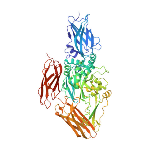
Crystal structure of transglutaminase 2 with GTP complex and amino acid sequence evidence of evolution of GTP binding site.

Jang, T.H.Lee, D.S.Choi, K.Jeong, E.M.Kim, I.G.Kim, Y.W.Chun, J.N.Jeon, J.H.Park, H.H.

(2014) PLoS One 9: e107005-e107005

  • DOI: https://doi.org/10.1371/journal.pone.0107005
  • Primary Citation Related Structures: 
    4PYG

  • PubMed Abstract: 

    Transglutaminase2 (TG2) is a multi-functional protein involved in various cellular processes, including apoptosis, differentiation, wound healing, and angiogenesis. The malfunction of TG2 causes many human disease including inflammatory disease, celiac disease, neurodegenerative diseases, tissue fibrosis, and cancers. Protein cross-linking activity, which is representative of TG2, is activated by calcium ions and suppressed by GTP. Here, we elucidated the structure of TG2 in complex with its endogenous inhibitor, GTP. Our structure showed why GTP is the optimal nucleotide for interacting with and inhibiting TG2. In addition, sequence comparison provided information describing the evolutionary scenario of GTP usage for controlling the activity of TG2.


  • Organizational Affiliation
    • School of Biotechnology, Yeungnam University, Gyeongsan, South Korea; Graduate School of Biochemistry, Yeungnam University, Gyeongsan, South Korea.

Macromolecule Content 

  • Total Structure Weight: 237.02 kDa 
  • Atom Count: 16,462 
  • Modeled Residue Count: 2,055 
  • Deposited Residue Count: 2,085 
  • Unique protein chains: 1

Macromolecules

Find similar proteins by:|  3D Structure
Entity ID: 1
MoleculeChains  Sequence LengthOrganismDetailsImage
Protein-glutamine gamma-glutamyltransferase 2A,
B,
C [auth E]
695Homo sapiensMutation(s): 0 
Gene Names: TGM2
EC: 2.3.2.13 (PDB Primary Data), 2.3.1 (UniProt), 3.4 (UniProt), 3.5.1.44 (UniProt)
UniProt & NIH Common Fund Data Resources
Find proteins for P21980 (Homo sapiens)
Explore P21980 
Go to UniProtKB:  P21980
PHAROS:  P21980
GTEx:  ENSG00000198959 
Entity Groups
Sequence Clusters30% Identity50% Identity70% Identity90% Identity95% Identity100% Identity
UniProt GroupP21980
Sequence Annotations
Expand
Reference Sequence

Experimental Data & Validation

Experimental Data

  • Method: X-RAY DIFFRACTION
  • Resolution: 2.80 Å
  • R-Value Free:  0.319 (Depositor), 0.309 (DCC) 
  • R-Value Work:  0.236 (Depositor), 0.230 (DCC) 
  • R-Value Observed: 0.240 (Depositor) 
Space Group: C 2 2 21
Unit Cell:
Length ( Å )Angle ( ˚ )
a = 133.546α = 90
b = 216.218β = 90
c = 165.183γ = 90
Software Package:
Software NamePurpose
HKL-2000data collection
PHASESphasing
REFMACrefinement
HKL-2000data reduction
HKL-2000data scaling

Structure Validation

View Full Validation Report


Ligand Structure Quality Assessment 


Entry History 

Deposition Data

Revision History  (Full details and data files)

  • Version 1.0: 2015-02-11
    Type: Initial release
  • Version 1.1: 2024-10-30
    Changes: Data collection, Database references, Derived calculations, Structure summary