


Crystal Structure of LysM domain from pteris ryukyuensis chitinase A
Ohnuma, T., Numata, T., Taira, T., Fukamizo, T.To be published.
Experimental Data Snapshot
wwPDB Validation 3D Report Full Report

Entity ID: 1 | |||||
|---|---|---|---|---|---|
| Molecule | Chains | Sequence Length | Organism | Details | Image |
| Chitinase A | 49 | Pteris ryukyuensis | Mutation(s): 0 Gene Names: prchiA | 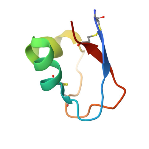 | |
UniProt | |||||
Entity Groups | |||||
| Sequence Clusters | 30% Identity50% Identity70% Identity90% Identity95% Identity100% Identity | ||||
| UniProt Group | Q0WYK2 | ||||
Sequence AnnotationsExpand | |||||
Reference Sequence | |||||
| Ligands 1 Unique | |||||
|---|---|---|---|---|---|
| ID | Chains | Name / Formula / InChI Key | 2D Diagram | 3D Interactions | |
| ZN Download:Ideal Coordinates CCD File | E [auth A], F [auth A], G [auth B], H [auth D] | ZINC ION Zn PTFCDOFLOPIGGS-UHFFFAOYSA-N |  | ||
| Length ( Å ) | Angle ( ˚ ) |
|---|---|
| a = 38.686 | α = 90 |
| b = 50.229 | β = 90 |
| c = 92.292 | γ = 90 |
| Software Name | Purpose |
|---|---|
| HKL-2000 | data collection |
| SnB | phasing |
| REFMAC | refinement |
| HKL-2000 | data reduction |
| HKL-2000 | data scaling |


