Crystal structures of influenza a virus matrix protein m1: variations on a theme.
Safo, M.K., Musayev, F.N., Mosier, P.D., Zhou, Q., Xie, H., Desai, U.R.(2014) PLoS One 9: e109510-e109510
- PubMed: 25295515 Search on PubMedSearch on PubMed Central
- DOI: https://doi.org/10.1371/journal.pone.0109510
- Primary Citation Related Structures:
4PUS - PubMed Abstract:
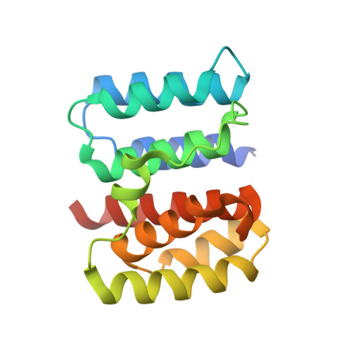
Matrix protein 1 (M1) of the influenza A virus plays multiple roles in virion assembly and infection. Interest in the pH dependence of M1's multiple functions led us to study the effect of subtle pH changes on M1 structure, resulting in the elucidation of a unique low-pH crystal structure of the N(1-165)-domain of A/WSN/33 (H1N1) M1 that has never been reported. Although the 2.2 Å crystal structure of M1 N-terminus shows a dimer with the two monomers interacting in a face-to-face fashion at low pH as observed earlier, a 44° rotation of the second monomer has led to a significantly different dimer interface that possibly affects dimer stability. More importantly, while one of the monomers is fully defined, the N-terminal half of the second monomer shows considerable disorder that appears inherent in the protein and is potentially physiologically relevant. Such disorder has not been observed in any other previously reported structure at either low or high pH conditions, despite similar crystallization pH conditions. By comparing our novel N(1-165)-domain structure with other low-pH or neutral-pH M1 structures, it appears that M1 can energetically access different monomer and dimer conformations, as well as oligomeric states, with varying degree of similarities. The study reported here provides further insights into M1 oligomerization that may be essential for viral propagation and infectivity.
- Department of Medicinal Chemistry, School of Pharmacy and Institute for Structural Biology and Drug Discovery, Virginia Commonwealth University, Richmond, Virginia, United States of America.
Organizational Affiliation: