4PTN

Crystal Structure of YagE, a KDG aldolase protein in complex with Magnesium cation coordinated L-glyceraldehyde


Experimental Data Snapshot

  • Method: X-RAY DIFFRACTION
  • Resolution: 1.99 Å
  • R-Value Free: 0.203 
  • R-Value Work: 0.174 
  • R-Value Observed: 0.176 

wwPDB Validation   3D Report Full Report


This is version 3.0 of the entry. See complete history


Literature

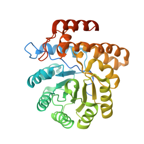
Crystal structure of YagE, a putative DHDPS-like protein from Escherichia coli K12.

Manicka, S.Peleg, Y.Unger, T.Albeck, S.Dym, O.Greenblatt, H.M.Bourenkov, G.Lamzin, V.Krishnaswamy, S.Sussman, J.L.

(2008) Proteins 71: 2102-2108


Macromolecules
Find similar proteins by:  (by identity cutoff)  |  3D Structure
Entity ID: 1
MoleculeChains Sequence LengthOrganismDetailsImage
Probable 2-keto-3-deoxy-galactonate aldolase YagE
A, B, C, D
343Escherichia coli K-12Mutation(s): 0 
Gene Names: b0268JW0261yagE
EC: 4.1.2
UniProt
Find proteins for P75682 (Escherichia coli (strain K12))
Explore P75682 
Go to UniProtKB:  P75682
Entity Groups  
Sequence Clusters30% Identity50% Identity70% Identity90% Identity95% Identity100% Identity
UniProt GroupP75682
Sequence Annotations
Expand
  • Reference Sequence
Small Molecules
Ligands 5 Unique
IDChains Name / Formula / InChI Key2D Diagram3D Interactions
GOL
Query on GOL

Download Ideal Coordinates CCD File 
M [auth B]GLYCEROL
C3 H8 O3
PEDCQBHIVMGVHV-UHFFFAOYSA-N
GXV
Query on GXV

Download Ideal Coordinates CCD File 
G [auth A],
N [auth B],
T [auth C],
X [auth D]
L-glyceraldehyde
C3 H6 O3
MNQZXJOMYWMBOU-GSVOUGTGSA-N
PYR
Query on PYR

Download Ideal Coordinates CCD File 
P [auth B],
Z [auth D]
PYRUVIC ACID
C3 H4 O3
LCTONWCANYUPML-UHFFFAOYSA-N
EDO
Query on EDO

Download Ideal Coordinates CCD File 
E [auth A]
F [auth A]
I [auth B]
J [auth B]
K [auth B]
E [auth A],
F [auth A],
I [auth B],
J [auth B],
K [auth B],
L [auth B],
Q [auth C],
R [auth C],
S [auth C],
V [auth D],
W [auth D]
1,2-ETHANEDIOL
C2 H6 O2
LYCAIKOWRPUZTN-UHFFFAOYSA-N
MG
Query on MG

Download Ideal Coordinates CCD File 
H [auth A],
O [auth B],
U [auth C],
Y [auth D]
MAGNESIUM ION
Mg
JLVVSXFLKOJNIY-UHFFFAOYSA-N
Experimental Data & Validation

Experimental Data

  • Method: X-RAY DIFFRACTION
  • Resolution: 1.99 Å
  • R-Value Free: 0.203 
  • R-Value Work: 0.174 
  • R-Value Observed: 0.176 
  • Space Group: P 21 21 2
Unit Cell:
Length ( Å )Angle ( ˚ )
a = 141.2α = 90
b = 155.07β = 90
c = 55.4γ = 90
Software Package:
Software NamePurpose
XSCALEdata scaling
PHASERphasing
REFMACrefinement
PDB_EXTRACTdata extraction

Structure Validation

View Full Validation Report


Entry History 

Deposition Data

Revision History  (Full details and data files)

  • Version 1.0: 2014-12-03
    Type: Initial release
  • Version 2.0: 2020-07-08
    Changes: Atomic model, Database references, Derived calculations, Structure summary
  • Version 3.0: 2023-11-15
    Changes: Advisory, Atomic model, Data collection, Database references, Derived calculations, Refinement description