


Crystal Structure of Engineered Protein OR464.
Vorobiev, S., Parmeggiani, F., Seetharaman, J., Huang, P.-S., Janjua, H., Xiao, R., Maglaqui, M., Park, K., Everett, J.K., Acton, T.B., Baker, D., Montelione, G.T., Tong, L., Hunt, J.To be published.
Experimental Data Snapshot
Starting Model: experimental
View more details
wwPDB Validation 3D Report Full Report

Entity ID: 1 | |||||
|---|---|---|---|---|---|
| Molecule | Chains | Sequence Length | Organism | Details | Image |
| OR464 | 271 | synthetic construct | Mutation(s): 0 | 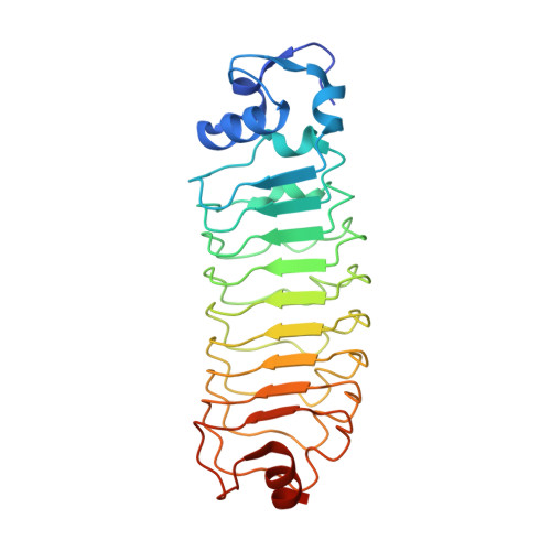 | |
| Length ( Å ) | Angle ( ˚ ) |
|---|---|
| a = 31.916 | α = 90 |
| b = 51.455 | β = 90.98 |
| c = 69.005 | γ = 90 |
| Software Name | Purpose |
|---|---|
| PHENIX | refinement |
| PDB_EXTRACT | data extraction |
| ADSC | data collection |
| HKL-2000 | data reduction |
| HKL-2000 | data scaling |
| BALBES | phasing |


