Structural studies on Mycobacterium tuberculosis RecA: Molecular plasticity and interspecies variability
Chandran, A.V., Prabu, J.R., Nautiyal, A., Patil, K.N., Muniyappa, K., Vijayan, M.(2015) J Biosci 40: 13-30
- PubMed: 25740138 Search on PubMed
- DOI: https://doi.org/10.1007/s12038-014-9497-x
- Primary Citation Related Structures:
4OQF, 4PO1, 4PO8, 4PO9, 4POA, 4PPF, 4PPG, 4PPN, 4PPQ, 4PQF, 4PQR, 4PQY, 4PR0, 4PSA, 4PSK, 4PSV, 4PTL - PubMed Abstract:
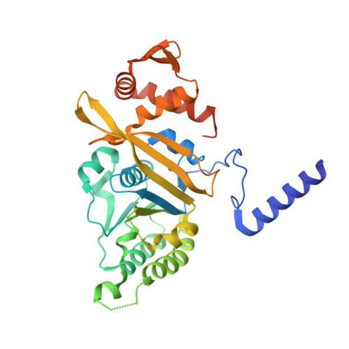
Structures of crystals of Mycobacterium tuberculosis RecA, grown and analysed under different conditions, provide insights into hitherto underappreciated details of molecular structure and plasticity. In particular, they yield information on the invariant and variable features of the geometry of the P-loop, whose binding to ATP is central for all the biochemical activities of RecA. The strengths of interaction of the ligands with the P-loop reveal significant differences. This in turn affects the magnitude of the motion of the 'switch' residue, Gln195 in M. tuberculosis RecA, which triggers the transmission of ATP-mediated allosteric information to the DNA binding region. M. tuberculosis RecA is substantially rigid compared with its counterparts from M. smegmatis and E. coli, which exhibit concerted internal molecular mobility. The interspecies variability in the plasticity of the two mycobacterial proteins is particularly surprising as they have similar sequence and 3D structure. Details of the interactions of ligands with the protein, characterized in the structures reported here, could be useful for design of inhibitors against M. tuberculosis RecA.
- Molecular Biophysics Unit, Indian Institute of Science, Bangalore 560 012.
Organizational Affiliation: