4PK6

Crystal structure of the indoleamine 2,3-dioxygenagse 1 (IDO1) complexed with imidazothiazole derivative


Experimental Data Snapshot

  • Method: X-RAY DIFFRACTION
  • Resolution: 3.45 Å
  • R-Value Free: 0.256 
  • R-Value Work: 0.190 
  • R-Value Observed: 0.193 

wwPDB Validation   3D Report Full Report


Ligand Structure Quality Assessment 


This is version 1.2 of the entry. See complete history


Literature

Crystal Structures and Structure-Activity Relationships of Imidazothiazole Derivatives as IDO1 Inhibitors.

Tojo, S.Kohno, T.Tanaka, T.Kamioka, S.Ota, Y.Ishii, T.Kamimoto, K.Asano, S.Isobe, Y.

(2014) ACS Med Chem Lett 5: 1119-1123

  • DOI: https://doi.org/10.1021/ml500247w
  • Primary Citation of Related Structures:  
    4PK5, 4PK6

  • PubMed Abstract: 

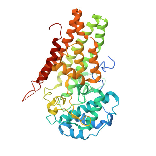
    Indoleamine 2,3-dioxygenase 1 (IDO1) is considered as a promising target for the treatment of several diseases, including neurological disorders and cancer. We report here the crystal structures of two IDO1/IDO1 inhibitor complexes, one of which shows that Amg-1 is directly bound to the heme iron of IDO1 with a clear induced fit. We also describe the identification and preliminary optimization of imidazothiazole derivatives as novel IDO1 inhibitors. Using our crystal structure information and structure-activity relationships (SAR) at the pocket-B of IDO1, we found a series of urea derivatives as potent IDO1 inhibitors and revealed that generation of an induced fit and the resulting interaction with Phe226 and Arg231 are essential for potent IDO1 inhibitory activity. The results of this study are very valuable for understanding the mechanism of IDO1 activation, which is very important for structure-based drug design (SBDD) to discover potent IDO1 inhibitors.


  • Organizational Affiliation

    Dainippon Sumitomo Pharma Co., Ltd. , 3-1-98 Kasugade-naka, Konohana-ku, Osaka 554-0022, Japan.


Macromolecules
Find similar proteins by:  (by identity cutoff)  |  3D Structure
Entity ID: 1
MoleculeChains Sequence LengthOrganismDetailsImage
Indoleamine 2,3-dioxygenase 1
A, B
423Homo sapiensMutation(s): 0 
Gene Names: IDO1IDOINDO
EC: 1.13.11.52
UniProt & NIH Common Fund Data Resources
Find proteins for P14902 (Homo sapiens)
Explore P14902 
Go to UniProtKB:  P14902
PHAROS:  P14902
GTEx:  ENSG00000131203 
Entity Groups  
Sequence Clusters30% Identity50% Identity70% Identity90% Identity95% Identity100% Identity
UniProt GroupP14902
Sequence Annotations
Expand
  • Reference Sequence
Small Molecules
Binding Affinity Annotations 
IDSourceBinding Affinity
PKL BindingDB:  4PK6 IC50: 1900 (nM) from 1 assay(s)
Experimental Data & Validation

Experimental Data

  • Method: X-RAY DIFFRACTION
  • Resolution: 3.45 Å
  • R-Value Free: 0.256 
  • R-Value Work: 0.190 
  • R-Value Observed: 0.193 
  • Space Group: P 21 21 21
Unit Cell:
Length ( Å )Angle ( ˚ )
a = 84.08α = 90
b = 91.12β = 90
c = 135.69γ = 90
Software Package:
Software NamePurpose
REFMACrefinement

Structure Validation

View Full Validation Report


Ligand Structure Quality Assessment 


Entry History 

Deposition Data

Revision History  (Full details and data files)

  • Version 1.0: 2014-09-03
    Type: Initial release
  • Version 1.1: 2014-10-29
    Changes: Database references
  • Version 1.2: 2017-10-04
    Changes: Data collection, Derived calculations, Other, Source and taxonomy