4PJG

Structure of human MR1-Ac-6-FP in complex with human MAIT B-F3-C1 TCR


Experimental Data Snapshot

  • Method: X-RAY DIFFRACTION
  • Resolution: 2.40 Å
  • R-Value Free: 0.213 
  • R-Value Work: 0.167 
  • R-Value Observed: 0.169 

wwPDB Validation   3D Report Full Report


Ligand Structure Quality Assessment 


This is version 1.3 of the entry. See complete history


Literature

A molecular basis underpinning the T cell receptor heterogeneity of mucosal-associated invariant T cells.

Eckle, S.B.Birkinshaw, R.W.Kostenko, L.Corbett, A.J.McWilliam, H.E.Reantragoon, R.Chen, Z.Gherardin, N.A.Beddoe, T.Liu, L.Patel, O.Meehan, B.Fairlie, D.P.Villadangos, J.A.Godfrey, D.I.Kjer-Nielsen, L.McCluskey, J.Rossjohn, J.

(2014) J Exp Med 211: 1585-1600

  • DOI: https://doi.org/10.1084/jem.20140484
  • Primary Citation of Related Structures:  
    4PJ5, 4PJ7, 4PJ8, 4PJ9, 4PJA, 4PJB, 4PJC, 4PJD, 4PJE, 4PJF, 4PJG, 4PJH, 4PJI, 4PJX

  • PubMed Abstract: 

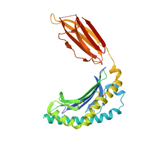
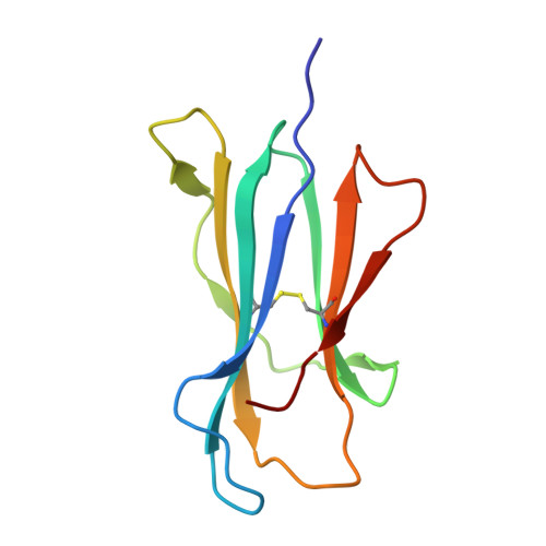
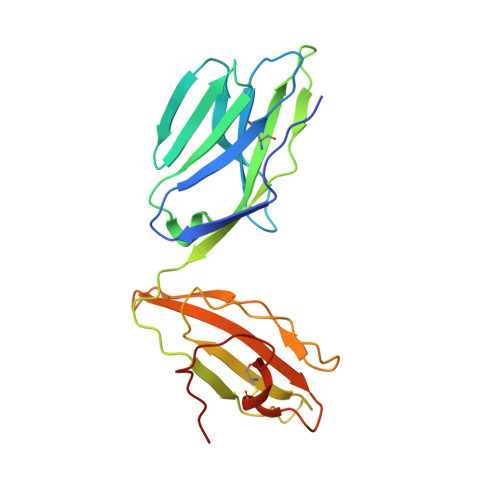
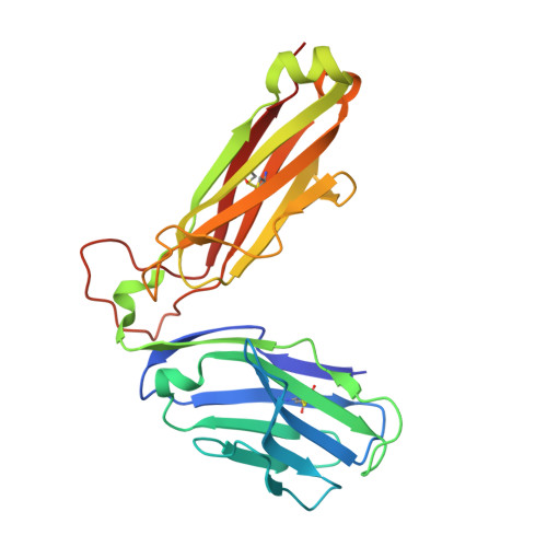
    Mucosal-associated invariant T (MAIT) cells express an invariant T cell receptor (TCR) α-chain (TRAV1-2 joined to TRAJ33, TRAJ20, or TRAJ12 in humans), which pairs with an array of TCR β-chains. MAIT TCRs can bind folate- and riboflavin-based metabolites restricted by the major histocompatibility complex (MHC)-related class I-like molecule, MR1. However, the impact of MAIT TCR and MR1-ligand heterogeneity on MAIT cell biology is unclear. We show how a previously uncharacterized MR1 ligand, acetyl-6-formylpterin (Ac-6-FP), markedly stabilized MR1, potently up-regulated MR1 cell surface expression, and inhibited MAIT cell activation. These enhanced properties of Ac-6-FP were attributable to structural alterations in MR1 that subsequently affected MAIT TCR recognition via conformational changes within the complementarity-determining region (CDR) 3β loop. Analysis of seven TRBV6-1(+) MAIT TCRs demonstrated how CDR3β hypervariability impacted on MAIT TCR recognition by altering TCR flexibility and contacts with MR1 and the Ag itself. Ternary structures of TRBV6-1, TRBV6-4, and TRBV20(+) MAIT TCRs in complex with MR1 bound to a potent riboflavin-based antigen (Ag) showed how variations in TRBV gene usage exclusively impacted on MR1 contacts within a consensus MAIT TCR-MR1 footprint. Moreover, differential TRAJ gene usage was readily accommodated within a conserved MAIT TCR-MR1-Ag docking mode. Collectively, MAIT TCR heterogeneity can fine-tune MR1 recognition in an Ag-dependent manner, thereby modulating MAIT cell recognition.


  • Organizational Affiliation

    Department of Microbiology and Immunology, Peter Doherty Institute for Infection and Immunity, University of Melbourne, Parkville, Victoria 3010, Australia.


Macromolecules
Find similar proteins by:  (by identity cutoff)  |  3D Structure
Entity ID: 1
MoleculeChains Sequence LengthOrganismDetailsImage
Major histocompatibility complex class I-related gene protein
A, C
271Homo sapiensMutation(s): 1 
Gene Names: MR1
UniProt & NIH Common Fund Data Resources
Find proteins for Q95460 (Homo sapiens)
Explore Q95460 
Go to UniProtKB:  Q95460
PHAROS:  Q95460
GTEx:  ENSG00000153029 
Entity Groups  
Sequence Clusters30% Identity50% Identity70% Identity90% Identity95% Identity100% Identity
UniProt GroupQ95460
Sequence Annotations
Expand
  • Reference Sequence
Find similar proteins by:  (by identity cutoff)  |  3D Structure
Entity ID: 2
MoleculeChains Sequence LengthOrganismDetailsImage
Beta-2-microglobulin
B, D
100Homo sapiensMutation(s): 0 
Gene Names: B2MCDABP0092HDCMA22P
UniProt & NIH Common Fund Data Resources
Find proteins for P61769 (Homo sapiens)
Explore P61769 
Go to UniProtKB:  P61769
PHAROS:  P61769
GTEx:  ENSG00000166710 
Entity Groups  
Sequence Clusters30% Identity50% Identity70% Identity90% Identity95% Identity100% Identity
UniProt GroupP61769
Sequence Annotations
Expand
  • Reference Sequence
Find similar proteins by:  (by identity cutoff)  |  3D Structure
Entity ID: 3
MoleculeChains Sequence LengthOrganismDetailsImage
TCR-alpha
E, G
205Homo sapiensMutation(s): 0 
Entity Groups  
Sequence Clusters30% Identity50% Identity70% Identity90% Identity95% Identity100% Identity
Sequence Annotations
Expand
  • Reference Sequence
Find similar proteins by:  (by identity cutoff)  |  3D Structure
Entity ID: 4
MoleculeChains Sequence LengthOrganismDetailsImage
TCR-beta
F, H
246Homo sapiensMutation(s): 0 
Entity Groups  
Sequence Clusters30% Identity50% Identity70% Identity90% Identity95% Identity100% Identity
Sequence Annotations
Expand
  • Reference Sequence
Small Molecules
Ligands 1 Unique
IDChains Name / Formula / InChI Key2D Diagram3D Interactions
30W
Query on 30W

Download Ideal Coordinates CCD File 
I [auth A],
J [auth C]
N-(6-formyl-4-oxo-3,4-dihydropteridin-2-yl)acetamide
C9 H7 N5 O3
DDBCPKAHJKOGKK-UHFFFAOYSA-N
Experimental Data & Validation

Experimental Data

  • Method: X-RAY DIFFRACTION
  • Resolution: 2.40 Å
  • R-Value Free: 0.213 
  • R-Value Work: 0.167 
  • R-Value Observed: 0.169 
  • Space Group: C 1 2 1
Unit Cell:
Length ( Å )Angle ( ˚ )
a = 217.7α = 90
b = 70.98β = 103.93
c = 142.34γ = 90
Software Package:
Software NamePurpose
BUSTERrefinement

Structure Validation

View Full Validation Report


Ligand Structure Quality Assessment 


Entry History 

Deposition Data

Revision History  (Full details and data files)

  • Version 1.0: 2014-07-02
    Type: Initial release
  • Version 1.1: 2014-08-06
    Changes: Database references
  • Version 1.2: 2014-10-01
    Changes: Database references
  • Version 1.3: 2023-12-27
    Changes: Data collection, Database references, Derived calculations, Other, Refinement description, Source and taxonomy