4PGQ | pdb_00004pgq

Structure of human DNA polymerase beta complexed with G in the template base paired with incoming non-hydrolyzable TTP


Experimental Data Snapshot

  • Method: X-RAY DIFFRACTION
  • Resolution: 2.30 Å
  • R-Value Free: 
    0.260 (Depositor), 0.260 (DCC) 
  • R-Value Work: 
    0.203 (Depositor), 0.205 (DCC) 
  • R-Value Observed: 
    0.206 (Depositor) 

wwPDB Validation 3D Report Full Report

Validation slider image for 4PGQ

Ligand Structure Quality Assessment 


This is version 1.4 of the entry. See complete history

Literature

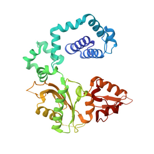
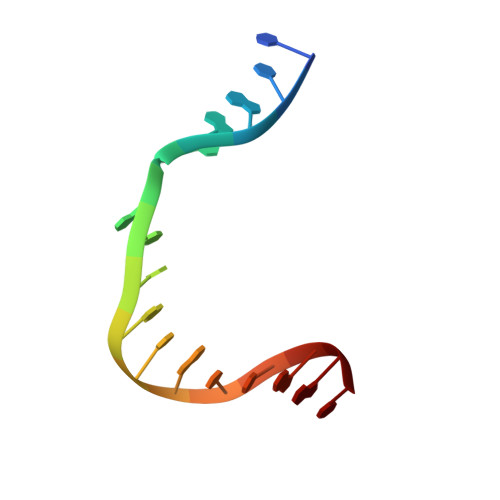
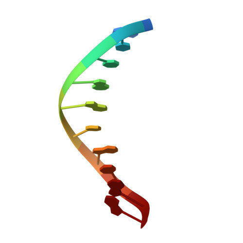
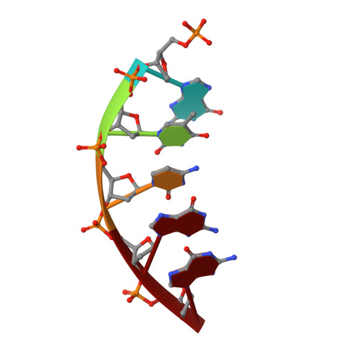
The spontaneous replication error and the mismatch discrimination mechanisms of human DNA polymerase beta.

Koag, M.C.Nam, K.Lee, S.

(2015) Nucleic Acids Res 42: 11233-11245

  • DOI: https://doi.org/10.1093/nar/gku789
  • Primary Citation Related Structures: 
    4PGQ, 4PGX, 4PHA, 4PHD

  • PubMed Abstract: 

    To provide molecular-level insights into the spontaneous replication error and the mismatch discrimination mechanisms of human DNA polymerase β (polβ), we report four crystal structures of polβ complexed with dG•dTTP and dA•dCTP mismatches in the presence of Mg2+ or Mn2+. The Mg(2+)-bound ground-state structures show that the dA•dCTP-Mg2+ complex adopts an 'intermediate' protein conformation while the dG•dTTP-Mg2+ complex adopts an open protein conformation. The Mn(2+)-bound 'pre-chemistry-state' structures show that the dA•dCTP-Mn2+ complex is structurally very similar to the dA•dCTP-Mg2+ complex, whereas the dG•dTTP-Mn2+ complex undergoes a large-scale conformational change to adopt a Watson-Crick-like dG•dTTP base pair and a closed protein conformation. These structural differences, together with our molecular dynamics simulation studies, suggest that polβ increases replication fidelity via a two-stage mismatch discrimination mechanism, where one is in the ground state and the other in the closed conformation state. In the closed conformation state, polβ appears to allow only a Watson-Crick-like conformation for purine•pyrimidine base pairs, thereby discriminating the mismatched base pairs based on their ability to form the Watson-Crick-like conformation. Overall, the present studies provide new insights into the spontaneous replication error and the replication fidelity mechanisms of polβ.


  • Organizational Affiliation
    • Division of Medicinal Chemistry, College of Pharmacy, The University of Texas at Austin, Austin, TX 78712, USA.

Macromolecule Content 

  • Total Structure Weight: 47.58 kDa 
  • Atom Count: 3,365 
  • Modeled Residue Count: 355 
  • Deposited Residue Count: 360 
  • Unique protein chains: 1
  • Unique nucleic acid chains: 3

Macromolecules


Find similar proteins by:|  3D Structure
Entity ID: 1
MoleculeChains  Sequence LengthOrganismDetailsImage
DNA polymerase beta329Homo sapiensMutation(s): 0 
Gene Names: POLB
EC: 2.7.7.7 (PDB Primary Data), 4.2.99 (PDB Primary Data), 4.2.99.18 (UniProt)
UniProt & NIH Common Fund Data Resources
Find proteins for P06746 (Homo sapiens)
Explore P06746 
Go to UniProtKB:  P06746
PHAROS:  P06746
GTEx:  ENSG00000070501 
Entity Groups
Sequence Clusters30% Identity50% Identity70% Identity90% Identity95% Identity100% Identity
UniProt GroupP06746
Sequence Annotations
Expand
Reference Sequence
Find similar nucleic acids by:  Sequence
Entity ID: 2
MoleculeChains LengthOrganismImage
DNA (5'-D(*CP*CP*GP*AP*CP*GP*TP*CP*GP*CP*AP*TP*CP*AP*GP*C)-3')B [auth T]16synthetic construct
Sequence Annotations
Expand
Reference Sequence
Find similar nucleic acids by:  Sequence
Entity ID: 3
MoleculeChains LengthOrganismImage
DNA (5'-D(*GP*CP*TP*GP*AP*TP*GP*CP*GP*A)-3')C [auth P]10synthetic construct
Sequence Annotations
Expand
Reference Sequence
Find similar nucleic acids by:  Sequence
Entity ID: 4
MoleculeChains LengthOrganismImage
DNA (5'-D(P*GP*TP*CP*GP*G)-3')5synthetic construct
Sequence Annotations
Expand
Reference Sequence

Experimental Data & Validation

Experimental Data

  • Method: X-RAY DIFFRACTION
  • Resolution: 2.30 Å
  • R-Value Free:  0.260 (Depositor), 0.260 (DCC) 
  • R-Value Work:  0.203 (Depositor), 0.205 (DCC) 
  • R-Value Observed: 0.206 (Depositor) 
Space Group: P 1 21 1
Unit Cell:
Length ( Å )Angle ( ˚ )
a = 54.898α = 90
b = 80.087β = 106.15
c = 55.077γ = 90
Software Package:
Software NamePurpose
PHENIXrefinement
DENZOdata reduction
PDB_EXTRACTdata extraction

Structure Validation

View Full Validation Report


Ligand Structure Quality Assessment 


Entry History 

& Funding Information

Deposition Data


Funding OrganizationLocationGrant Number
National Institutes of Health/National Institute of Environmental Health Sciences (NIH/NIEHS)United StatesES23101

Revision History  (Full details and data files)

  • Version 1.0: 2014-10-01
    Type: Initial release
  • Version 1.1: 2014-10-08
    Changes: Database references
  • Version 1.2: 2017-09-20
    Changes: Author supporting evidence, Database references, Derived calculations, Other, Refinement description, Source and taxonomy
  • Version 1.3: 2019-12-18
    Changes: Advisory, Author supporting evidence, Derived calculations
  • Version 1.4: 2023-12-27
    Changes: Data collection, Database references, Derived calculations, Refinement description