4PG8

Crystal structure of S. aureus Homoserine Dehydrogenase at pH8.5


Experimental Data Snapshot

  • Method: X-RAY DIFFRACTION
  • Resolution: 2.20 Å
  • R-Value Free: 0.237 
  • R-Value Work: 0.201 
  • R-Value Observed: 0.203 

wwPDB Validation   3D Report Full Report


This is version 1.4 of the entry. See complete history


Literature

Structural basis for the catalytic mechanism of homoserine dehydrogenase.

Navratna, V.Reddy, G.Gopal, B.

(2015) Acta Crystallogr D Biol Crystallogr 71: 1216-1225

  • DOI: https://doi.org/10.1107/S1399004715004617
  • Primary Citation of Related Structures:  
    4PG4, 4PG5, 4PG6, 4PG7, 4PG8

  • PubMed Abstract: 

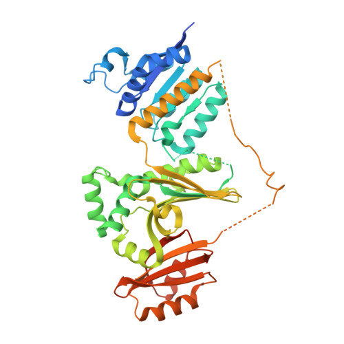
    Homoserine dehydrogenase (HSD) is an oxidoreductase in the aspartic acid pathway. This enzyme coordinates a critical branch point of the metabolic pathway that leads to the synthesis of bacterial cell-wall components such as L-lysine and m-DAP in addition to other amino acids such as L-threonine, L-methionine and L-isoleucine. Here, a structural rationale for the hydride-transfer step in the reaction mechanism of HSD is reported. The structure of Staphylococcus aureus HSD was determined at different pH conditions to understand the basis for the enhanced enzymatic activity at basic pH. An analysis of the crystal structure revealed that Lys105, which is located at the interface of the catalytic and cofactor-binding sites, could mediate the hydride-transfer step of the reaction mechanism. The role of Lys105 was subsequently confirmed by mutational analysis. Put together, these studies reveal the role of conserved water molecules and a lysine residue in hydride transfer between the substrate and the cofactor.


  • Organizational Affiliation

    Molecular Biophysics Unit, Indian Institute of Science, Bangalore, Karnataka 560 012, India.


Macromolecules
Find similar proteins by:  (by identity cutoff)  |  3D Structure
Entity ID: 1
MoleculeChains Sequence LengthOrganismDetailsImage
Homoserine dehydrogenase
A, B
468Staphylococcus aureus M1064Mutation(s): 0 
Gene Names: U5K_01898
EC: 1.1.1.3
UniProt
Find proteins for A0A0M3KKV6 (Staphylococcus aureus M1064)
Explore A0A0M3KKV6 
Go to UniProtKB:  A0A0M3KKV6
Entity Groups  
Sequence Clusters30% Identity50% Identity70% Identity90% Identity95% Identity100% Identity
UniProt GroupA0A0M3KKV6
Sequence Annotations
Expand
  • Reference Sequence
Small Molecules
Ligands 4 Unique
IDChains Name / Formula / InChI Key2D Diagram3D Interactions
PEG
Query on PEG

Download Ideal Coordinates CCD File 
L [auth A],
U [auth B],
V [auth B]
DI(HYDROXYETHYL)ETHER
C4 H10 O3
MTHSVFCYNBDYFN-UHFFFAOYSA-N
GOL
Query on GOL

Download Ideal Coordinates CCD File 
T [auth B]GLYCEROL
C3 H8 O3
PEDCQBHIVMGVHV-UHFFFAOYSA-N
DMS
Query on DMS

Download Ideal Coordinates CCD File 
W [auth B]DIMETHYL SULFOXIDE
C2 H6 O S
IAZDPXIOMUYVGZ-UHFFFAOYSA-N
ACT
Query on ACT

Download Ideal Coordinates CCD File 
C [auth A]
D [auth A]
E [auth A]
F [auth A]
G [auth A]
C [auth A],
D [auth A],
E [auth A],
F [auth A],
G [auth A],
H [auth A],
I [auth A],
J [auth A],
K [auth A],
M [auth B],
N [auth B],
O [auth B],
P [auth B],
Q [auth B],
R [auth B],
S [auth B]
ACETATE ION
C2 H3 O2
QTBSBXVTEAMEQO-UHFFFAOYSA-M
Experimental Data & Validation

Experimental Data

  • Method: X-RAY DIFFRACTION
  • Resolution: 2.20 Å
  • R-Value Free: 0.237 
  • R-Value Work: 0.201 
  • R-Value Observed: 0.203 
  • Space Group: P 21 21 21
Unit Cell:
Length ( Å )Angle ( ˚ )
a = 72.87α = 90
b = 117.51β = 90
c = 119.53γ = 90
Software Package:
Software NamePurpose
HKL-3000data collection
iMOSFLMdata reduction
SCALAdata scaling
PHASERphasing
Cootmodel building
REFMACrefinement
PDB_EXTRACTdata extraction

Structure Validation

View Full Validation Report


Entry History 

Deposition Data

Revision History  (Full details and data files)

  • Version 1.0: 2015-05-06
    Type: Initial release
  • Version 1.1: 2015-05-13
    Changes: Database references
  • Version 1.2: 2015-07-01
    Changes: Database references
  • Version 1.3: 2017-09-27
    Changes: Author supporting evidence, Derived calculations, Other, Refinement description, Source and taxonomy
  • Version 1.4: 2023-12-27
    Changes: Data collection, Database references, Refinement description