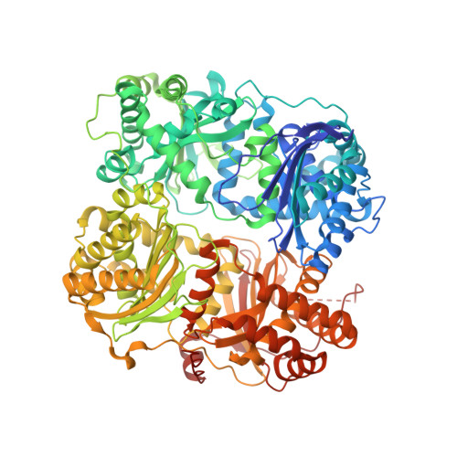
Dual Exosite-binding Inhibitors of Insulin-degrading Enzyme Challenge Its Role as the Primary Mediator of Insulin Clearance in Vivo.
Durham, T.B., Toth, J.L., Klimkowski, V.J., Cao, J.X., Siesky, A.M., Alexander-Chacko, J., Wu, G.Y., Dixon, J.T., McGee, J.E., Wang, Y., Guo, S.Y., Cavitt, R.N., Schindler, J., Thibodeaux, S.J., Calvert, N.A., Coghlan, M.J., Sindelar, D.K., Christe, M., Kiselyov, V.V., Michael, M.D., Sloop, K.W.(2015) J Biological Chem 290: 20044-20059
- PubMed: 26085101
- DOI: https://doi.org/10.1074/jbc.M115.638205
- Primary Citation of Related Structures:
4PF7, 4PF9, 4PFC - PubMed Abstract:
Insulin-degrading enzyme (IDE, insulysin) is the best characterized catabolic enzyme implicated in proteolysis of insulin. Recently, a peptide inhibitor of IDE has been shown to affect levels of insulin, amylin, and glucagon in vivo. However, IDE(-/-) mice display variable phenotypes relating to fasting plasma insulin levels, glucose tolerance, and insulin sensitivity depending on the cohort and age of animals. Here, we interrogated the importance of IDE-mediated catabolism on insulin clearance in vivo. Using a structure-based design, we linked two newly identified ligands binding at unique IDE exosites together to construct a potent series of novel inhibitors. These compounds do not interact with the catalytic zinc of the protease. Because one of these inhibitors (NTE-1) was determined to have pharmacokinetic properties sufficient to sustain plasma levels >50 times its IDE IC50 value, studies in rodents were conducted. In oral glucose tolerance tests with diet-induced obese mice, NTE-1 treatment improved the glucose excursion. Yet in insulin tolerance tests and euglycemic clamp experiments, NTE-1 did not enhance insulin action or increase plasma insulin levels. Importantly, IDE inhibition with NTE-1 did result in elevated plasma amylin levels, suggesting the in vivo role of IDE action on amylin may be more significant than an effect on insulin. Furthermore, using the inhibitors described in this report, we demonstrate that in HEK cells IDE has little impact on insulin clearance. In total, evidence from our studies supports a minimal role for IDE in insulin metabolism in vivo and suggests IDE may be more important in helping regulate amylin clearance.
- From the Discovery Chemistry Research and Technologies, durham_timothy_barrett@lilly.com.
Organizational Affiliation:


