4PDJ

Neutron crystal Structure of E.coli Dihydrofolate Reductase complexed with folate and NADP+


Experimental Data Snapshot

  • Method: X-RAY DIFFRACTION
  • Resolution: 1.60 Å
  • R-Value Free: 0.218 
  • R-Value Work: 0.194 
  • R-Value Observed: 0.195 

  • Method: NEUTRON DIFFRACTION
  • Resolution: 1.99 Å
  • R-Value Free: 0.271 
  • R-Value Work: 0.230 
  • R-Value Observed: 0.232 

wwPDB Validation   3D Report Full Report


This is version 1.5 of the entry. See complete history


Literature

Toward resolving the catalytic mechanism of dihydrofolate reductase using neutron and ultrahigh-resolution X-ray crystallography.

Wan, Q.Bennett, B.C.Wilson, M.A.Kovalevsky, A.Langan, P.Howell, E.E.Dealwis, C.

(2014) Proc Natl Acad Sci U S A 111: 18225-18230

  • DOI: https://doi.org/10.1073/pnas.1415856111
  • Primary Citation of Related Structures:  
    4PDJ, 4PSY, 4RGC

  • PubMed Abstract: 

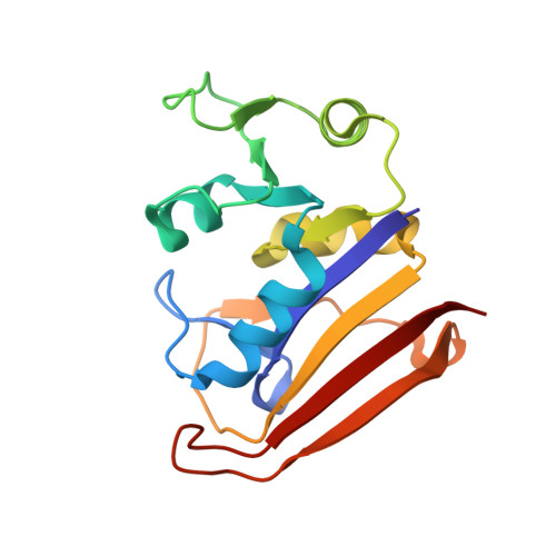
    Dihydrofolate reductase (DHFR) catalyzes the NADPH-dependent reduction of dihydrofolate (DHF) to tetrahydrofolate (THF). An important step in the mechanism involves proton donation to the N5 atom of DHF. The inability to determine the protonation states of active site residues and substrate has led to a lack of consensus regarding the catalytic mechanism involved. To resolve this ambiguity, we conducted neutron and ultrahigh-resolution X-ray crystallographic studies of the pseudo-Michaelis ternary complex of Escherichia coli DHFR with folate and NADP(+). The neutron data were collected to 2.0-Å resolution using a 3.6-mm(3) crystal with the quasi-Laue technique. The structure reveals that the N3 atom of folate is protonated, whereas Asp27 is negatively charged. Previous mechanisms have proposed a keto-to-enol tautomerization of the substrate to facilitate protonation of the N5 atom. The structure supports the existence of the keto tautomer owing to protonation of the N3 atom, suggesting that tautomerization is unnecessary for catalysis. In the 1.05-Å resolution X-ray structure of the ternary complex, conformational disorder of the Met20 side chain is coupled to electron density for a partially occupied water within hydrogen-bonding distance of the N5 atom of folate; this suggests direct protonation of substrate by solvent. We propose a catalytic mechanism for DHFR that involves stabilization of the keto tautomer of the substrate, elevation of the pKa value of the N5 atom of DHF by Asp27, and protonation of N5 by water that gains access to the active site through fluctuation of the Met20 side chain even though the Met20 loop is closed.


  • Organizational Affiliation

    Jiangsu Key Laboratory of Integrated Traditional Chinese and Western Medicine for Prevention and Treatment of Senile Disease, Yangzhou 225001, People's Republic of China; Jiangsu Co-Innovation Center for Prevention and Control of Important Animal Infectious Disease and Zoonoses, Yangzhou 225009, People's Republic of China; Department of Biochemistry, College of Medicine, Yangzhou University, Yangzhou 225001, People's Republic of China;


Macromolecules
Find similar proteins by:  (by identity cutoff)  |  3D Structure
Entity ID: 1
MoleculeChains Sequence LengthOrganismDetailsImage
Dihydrofolate reductase159Escherichia coli K-12Mutation(s): 0 
Gene Names: folAtmrAb0048JW0047
EC: 1.5.1.3
UniProt
Find proteins for P0ABQ4 (Escherichia coli (strain K12))
Explore P0ABQ4 
Go to UniProtKB:  P0ABQ4
Entity Groups  
Sequence Clusters30% Identity50% Identity70% Identity90% Identity95% Identity100% Identity
UniProt GroupP0ABQ4
Sequence Annotations
Expand
  • Reference Sequence
Small Molecules
Ligands 3 Unique
IDChains Name / Formula / InChI Key2D Diagram3D Interactions
NDP
Query on NDP

Download Ideal Coordinates CCD File 
E [auth A]NADPH DIHYDRO-NICOTINAMIDE-ADENINE-DINUCLEOTIDE PHOSPHATE
C21 H30 N7 O17 P3
ACFIXJIJDZMPPO-NNYOXOHSSA-N
DHF
Query on DHF

Download Ideal Coordinates CCD File 
D [auth A]DIHYDROFOLIC ACID
C19 H21 N7 O6
OZRNSSUDZOLUSN-LBPRGKRZSA-N
MN
Query on MN

Download Ideal Coordinates CCD File 
B [auth A],
C [auth A]
MANGANESE (II) ION
Mn
WAEMQWOKJMHJLA-UHFFFAOYSA-N
Experimental Data & Validation

Experimental Data

  • Method: X-RAY DIFFRACTION
  • Resolution: 1.60 Å
  • R-Value Free: 0.218 
  • R-Value Work: 0.194 
  • R-Value Observed: 0.195 
  • Space Group: P 21 21 21
  • Method: NEUTRON DIFFRACTION
  • Resolution: 1.99 Å
  • R-Value Free: 0.271 
  • R-Value Work: 0.230 
  • R-Value Observed: 0.232 
  • Space Group: P 21 21 21
Unit Cell:
Length ( Å )Angle ( ˚ )
a = 34.293α = 90
b = 45.625β = 90
c = 98.974γ = 90
Software Package:
Software NamePurpose
PHENIXrefinement
HKL-3000data processing
Cootmodel building
PDB_EXTRACTdata extraction
Cootmodel building
SCALAdata reduction
PHASERphasing
SCALAdata scaling
LAUEGENdata reduction

Structure Validation

View Full Validation Report


Entry History & Funding Information

Deposition Data


Funding OrganizationLocationGrant Number
Department of Energy (DOE, United States)United StatesFWP ERKP752
National Institutes of Health/National Institute of General Medical Sciences (NIH/NIGMS)United StatesR01GM071939
National Institutes of Health/National Institute of General Medical Sciences (NIH/NIGMS)United StatesR01GM092999
Yangzhou UniversityChina2013CXJ083
State Education MinistryChina--

Revision History  (Full details and data files)

  • Version 1.0: 2015-04-15
    Type: Initial release
  • Version 1.1: 2016-07-20
    Changes: Data collection
  • Version 1.2: 2017-09-20
    Changes: Author supporting evidence, Derived calculations, Source and taxonomy
  • Version 1.3: 2019-12-04
    Changes: Author supporting evidence
  • Version 1.4: 2023-12-27
    Changes: Data collection, Database references, Derived calculations
  • Version 1.5: 2024-04-03
    Changes: Refinement description