4P9C

Crystal structure of dCMP deaminase from the cyanophage S-TIM5 in complex with dCMP and dUMP


Experimental Data Snapshot

  • Method: X-RAY DIFFRACTION
  • Resolution: 2.60 Å
  • R-Value Free: 0.268 
  • R-Value Work: 0.231 
  • R-Value Observed: 0.233 

wwPDB Validation   3D Report Full Report


Ligand Structure Quality Assessment 


This is version 1.4 of the entry. See complete history


Literature

The First Crystal Structure of a dTTP-bound Deoxycytidylate Deaminase Validates and Details the Allosteric-Inhibitor Binding Site.

Marx, A.Alian, A.

(2015) J Biol Chem 290: 682-690

  • DOI: https://doi.org/10.1074/jbc.M114.617720
  • Primary Citation of Related Structures:  
    4P9C, 4P9D, 4P9E

  • PubMed Abstract: 

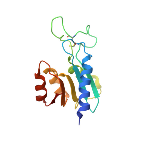
    Deoxycytidylate deaminase is unique within the zinc-dependent cytidine deaminase family as being allosterically regulated, activated by dCTP, and inhibited by dTTP. Here we present the first crystal structure of a dTTP-bound deoxycytidylate deaminase from the bacteriophage S-TIM5, confirming that this inhibitor binds to the same site as the dCTP activator. The molecular details of this structure, complemented by structures apo- and dCMP-bound, provide insights into the allosteric mechanism. Although the positioning of the nucleoside moiety of dTTP is almost identical to that previously described for dCTP, protonation of N3 in deoxythymidine and not deoxycytidine would facilitate hydrogen bonding of dTTP but not dCTP and may result in a higher affinity of dTTP to the allosteric site conferring its inhibitory activity. Further the functional group on C4 (O in dTTP and NH2 in dCTP) makes interactions with nonconserved protein residues preceding the allosteric motif, and the relative strength of binding to these residues appears to correspond to the potency of dTTP inhibition. The active sites of these structures are also uniquely occupied by dTMP and dCMP resolving aspects of substrate specificity. The methyl group of dTMP apparently clashes with a highly conserved tyrosine residue, preventing the formation of a correct base stacking shown to be imperative for deamination activity. The relevance of these findings to the wider zinc-dependent cytidine deaminase family is also discussed.


  • Organizational Affiliation

    From the Faculty of Biology, Technion-Israel Institute of Technology, Haifa 320003, Israel.


Macromolecules
Find similar proteins by:  (by identity cutoff)  |  3D Structure
Entity ID: 1
MoleculeChains Sequence LengthOrganismDetailsImage
Deoxycytidylate deaminase
A, B, C, D, E
A, B, C, D, E, F, G, H, I, J, K, L
138Cyanophage S-TIM5Mutation(s): 0 
UniProt
Find proteins for H6WFU3 (Cyanophage S-TIM5)
Explore H6WFU3 
Go to UniProtKB:  H6WFU3
Entity Groups  
Sequence Clusters30% Identity50% Identity70% Identity90% Identity95% Identity100% Identity
UniProt GroupH6WFU3
Sequence Annotations
Expand
  • Reference Sequence
Small Molecules
Ligands 3 Unique
IDChains Name / Formula / InChI Key2D Diagram3D Interactions
DU
Query on DU

Download Ideal Coordinates CCD File 
AA [auth E]
DA [auth F]
GA [auth G]
JA [auth H]
MA [auth I]
AA [auth E],
DA [auth F],
GA [auth G],
JA [auth H],
MA [auth I],
O [auth A],
PA [auth J],
R [auth B],
SA [auth K],
U [auth C],
VA [auth L],
X [auth D]
2'-DEOXYURIDINE-5'-MONOPHOSPHATE
C9 H13 N2 O8 P
JSRLJPSBLDHEIO-SHYZEUOFSA-N
DCM
Query on DCM

Download Ideal Coordinates CCD File 
CA [auth F]
FA [auth G]
IA [auth H]
LA [auth I]
N [auth A]
CA [auth F],
FA [auth G],
IA [auth H],
LA [auth I],
N [auth A],
OA [auth J],
Q [auth B],
RA [auth K],
T [auth C],
UA [auth L],
W [auth D],
Z [auth E]
2'-DEOXYCYTIDINE-5'-MONOPHOSPHATE
C9 H14 N3 O7 P
NCMVOABPESMRCP-SHYZEUOFSA-N
ZN
Query on ZN

Download Ideal Coordinates CCD File 
BA [auth F]
EA [auth G]
HA [auth H]
KA [auth I]
M [auth A]
BA [auth F],
EA [auth G],
HA [auth H],
KA [auth I],
M [auth A],
NA [auth J],
P [auth B],
QA [auth K],
S [auth C],
TA [auth L],
V [auth D],
Y [auth E]
ZINC ION
Zn
PTFCDOFLOPIGGS-UHFFFAOYSA-N
Experimental Data & Validation

Experimental Data

  • Method: X-RAY DIFFRACTION
  • Resolution: 2.60 Å
  • R-Value Free: 0.268 
  • R-Value Work: 0.231 
  • R-Value Observed: 0.233 
  • Space Group: C 1 2 1
Unit Cell:
Length ( Å )Angle ( ˚ )
a = 135.05α = 90
b = 147.21β = 95.62
c = 100.47γ = 90
Software Package:
Software NamePurpose
REFMACrefinement
SCALAdata scaling

Structure Validation

View Full Validation Report


Ligand Structure Quality Assessment 


Entry History 

Deposition Data

Revision History  (Full details and data files)

  • Version 1.0: 2014-11-26
    Type: Initial release
  • Version 1.1: 2014-12-03
    Changes: Database references
  • Version 1.2: 2015-01-14
    Changes: Database references
  • Version 1.3: 2017-11-22
    Changes: Database references, Derived calculations, Other, Refinement description, Source and taxonomy
  • Version 1.4: 2023-12-27
    Changes: Data collection, Database references, Derived calculations, Refinement description