4P79

Crystal structure of mouse claudin-15


Experimental Data Snapshot

  • Method: X-RAY DIFFRACTION
  • Resolution: 2.40 Å
  • R-Value Free: 0.253 
  • R-Value Work: 0.221 
  • R-Value Observed: 0.224 

wwPDB Validation   3D Report Full Report


Ligand Structure Quality Assessment 


This is version 1.4 of the entry. See complete history


Literature

Crystal structure of a claudin provides insight into the architecture of tight junctions.

Suzuki, H.Nishizawa, T.Tani, K.Yamazaki, Y.Tamura, A.Ishitani, R.Dohmae, N.Tsukita, S.Nureki, O.Fujiyoshi, Y.

(2014) Science 344: 304-307

  • DOI: https://doi.org/10.1126/science.1248571
  • Primary Citation of Related Structures:  
    4P79

  • PubMed Abstract: 

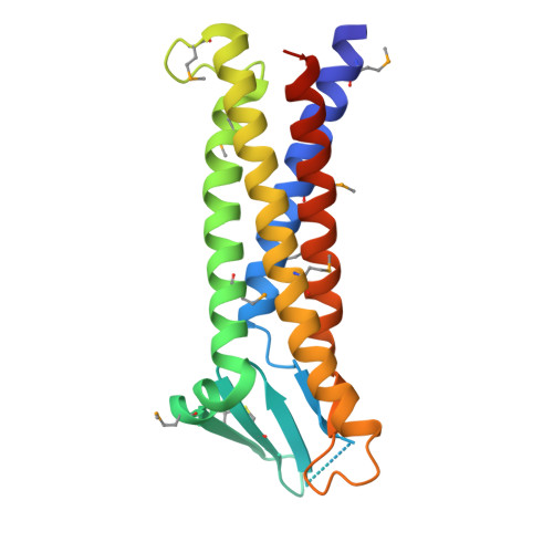
    Tight junctions are cell-cell adhesion structures in epithelial cell sheets that surround organ compartments in multicellular organisms and regulate the permeation of ions through the intercellular space. Claudins are the major constituents of tight junctions and form strands that mediate cell adhesion and function as paracellular barriers. We report the structure of mammalian claudin-15 at a resolution of 2.4 angstroms. The structure reveals a characteristic β-sheet fold comprising two extracellular segments, which is anchored to a transmembrane four-helix bundle by a consensus motif. Our analyses suggest potential paracellular pathways with distinctive charges on the extracellular surface, providing insight into the molecular basis of ion homeostasis across tight junctions.


  • Organizational Affiliation

    Cellular and Structural Physiology Institute, Nagoya University, Chikusa, Nagoya 464-8601, Japan.


Macromolecules
Find similar proteins by:  (by identity cutoff)  |  3D Structure
Entity ID: 1
MoleculeChains Sequence LengthOrganismDetailsImage
Claudin-15198Mus musculusMutation(s): 4 
Gene Names: Cldn15
Membrane Entity: Yes 
UniProt
Find proteins for Q9Z0S5 (Mus musculus)
Explore Q9Z0S5 
Go to UniProtKB:  Q9Z0S5
Entity Groups  
Sequence Clusters30% Identity50% Identity70% Identity90% Identity95% Identity100% Identity
UniProt GroupQ9Z0S5
Sequence Annotations
Expand
  • Reference Sequence
Small Molecules
Ligands 1 Unique
IDChains Name / Formula / InChI Key2D Diagram3D Interactions
OLC
Query on OLC

Download Ideal Coordinates CCD File 
B [auth A],
C [auth A]
(2R)-2,3-dihydroxypropyl (9Z)-octadec-9-enoate
C21 H40 O4
RZRNAYUHWVFMIP-GDCKJWNLSA-N
Modified Residues  1 Unique
IDChains TypeFormula2D DiagramParent
MSE
Query on MSE
A
L-PEPTIDE LINKINGC5 H11 N O2 SeMET
Experimental Data & Validation

Experimental Data

  • Method: X-RAY DIFFRACTION
  • Resolution: 2.40 Å
  • R-Value Free: 0.253 
  • R-Value Work: 0.221 
  • R-Value Observed: 0.224 
  • Space Group: C 1 2 1
Unit Cell:
Length ( Å )Angle ( ˚ )
a = 123.25α = 90
b = 28.403β = 100.84
c = 58.587γ = 90
Software Package:
Software NamePurpose
XDSdata scaling
PDB_EXTRACTdata extraction
PHENIXrefinement

Structure Validation

View Full Validation Report


Ligand Structure Quality Assessment 


Entry History 

Deposition Data

Revision History  (Full details and data files)

  • Version 1.0: 2014-04-30
    Type: Initial release
  • Version 1.1: 2014-05-07
    Changes: Derived calculations
  • Version 1.2: 2017-09-27
    Changes: Data collection, Database references, Derived calculations, Other, Refinement description, Source and taxonomy
  • Version 1.3: 2017-10-11
    Changes: Data collection
  • Version 1.4: 2023-12-27
    Changes: Data collection, Database references, Refinement description