4P6C

Structure of ribB complexed with inhibitor 4PEH


Experimental Data Snapshot

  • Method: X-RAY DIFFRACTION
  • Resolution: 1.86 Å
  • R-Value Free: 0.207 
  • R-Value Work: 0.163 
  • R-Value Observed: 0.165 

wwPDB Validation   3D Report Full Report


Ligand Structure Quality Assessment 


This is version 1.5 of the entry. See complete history


Literature

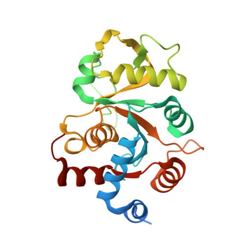
Structural Basis for Competitive Inhibition of 3,4-Dihydroxy-2-butanone-4-phosphate Synthase from Vibrio cholerae.

Islam, Z.Kumar, A.Singh, S.Salmon, L.Karthikeyan, S.

(2015) J Biol Chem 290: 11293-11308

  • DOI: https://doi.org/10.1074/jbc.M114.611830
  • Primary Citation of Related Structures:  
    4P6C, 4P6D, 4P6P, 4P77, 4P8E, 4P8J

  • PubMed Abstract: 

    The riboflavin biosynthesis pathway has been shown to be essential in many pathogens and is absent in humans. Therefore, enzymes involved in riboflavin synthesis are considered as potential antibacterial drug targets. The enzyme 3,4-dihydroxy-2-butanone-4-phosphate synthase (DHBPS) catalyzes one of the two committed steps in the riboflavin pathway and converts d-ribulose 5-phosphate (Ru5P) to l-3,4-dihydroxy-2-butanone 4-phosphate and formate. Moreover, DHBPS is shown to be indispensable for Mycobacterium, Salmonella, and Helicobacter species. Despite the essentiality of this enzyme in bacteria, no inhibitor has been identified hitherto. Here, we describe kinetic and crystal structure characterization of DHBPS from Vibrio cholerae (vDHBPS) with a competitive inhibitor 4-phospho-d-erythronohydroxamic acid (4PEH) at 1.86-Å resolution. In addition, we also report the structural characterization of vDHBPS in its apo form and in complex with its substrate and substrate plus metal ions at 1.96-, 1.59-, and 2.04-Å resolution, respectively. Comparison of these crystal structures suggests that 4PEH inhibits the catalytic activity of DHBPS as it is unable to form a proposed intermediate that is crucial for DHBPS activity. Furthermore, vDHBPS structures complexed with substrate and metal ions reveal that, unlike Candida albicans, binding of substrate to vDHBPS induces a conformational change from an open to closed conformation. Interestingly, the position of second metal ion, which is different from that of Methanococcus jannaschii, strongly supports an active role in the catalytic mechanism. Thus, the kinetic and structural characterization of vDHBPS reveals the molecular mechanism of inhibition shown by 4PEH and that it can be explored further for designing novel antibiotics.


  • Organizational Affiliation

    From the CSIR-Institute of Microbial Technology, Council of Scientific and Industrial Research (CSIR), Sector 39-A, Chandigarh 160 036, India and.


Macromolecules
Find similar proteins by:  (by identity cutoff)  |  3D Structure
Entity ID: 1
MoleculeChains Sequence LengthOrganismDetailsImage
3,4-dihydroxy-2-butanone 4-phosphate synthase
A, B
238Vibrio cholerae O1 biovar El Tor str. N16961Mutation(s): 0 
Gene Names: ribBVC_A1060
EC: 4.1.99.12
UniProt
Find proteins for Q9KKP2 (Vibrio cholerae serotype O1 (strain ATCC 39315 / El Tor Inaba N16961))
Explore Q9KKP2 
Go to UniProtKB:  Q9KKP2
Entity Groups  
Sequence Clusters30% Identity50% Identity70% Identity90% Identity95% Identity100% Identity
UniProt GroupQ9KKP2
Sequence Annotations
Expand
  • Reference Sequence
Small Molecules
Ligands 1 Unique
IDChains Name / Formula / InChI Key2D Diagram3D Interactions
RES
Query on RES

Download Ideal Coordinates CCD File 
C [auth A],
D [auth B]
4-PHOSPHO-D-ERYTHRONOHYDROXAMIC ACID
C4 H10 N O8 P
JJQQOJRGUHNREK-PWNYCUMCSA-N
Binding Affinity Annotations 
IDSourceBinding Affinity
RES Binding MOAD:  4P6C Ki: 1.00e+5 (nM) from 1 assay(s)
Experimental Data & Validation

Experimental Data

  • Method: X-RAY DIFFRACTION
  • Resolution: 1.86 Å
  • R-Value Free: 0.207 
  • R-Value Work: 0.163 
  • R-Value Observed: 0.165 
  • Space Group: P 21 21 21
Unit Cell:
Length ( Å )Angle ( ˚ )
a = 52.674α = 90
b = 72.654β = 90
c = 100.085γ = 90
Software Package:
Software NamePurpose
PHENIXrefinement
MAR345dtbdata collection
XDSdata scaling
PHASERphasing

Structure Validation

View Full Validation Report


Ligand Structure Quality Assessment 


Entry History & Funding Information

Deposition Data


Funding OrganizationLocationGrant Number
CSIRIndiaNWP104, NWP210

Revision History  (Full details and data files)

  • Version 1.0: 2015-03-25
    Type: Initial release
  • Version 1.1: 2015-04-01
    Changes: Database references
  • Version 1.2: 2015-04-29
    Changes: Structure summary
  • Version 1.3: 2015-05-13
    Changes: Database references
  • Version 1.4: 2017-11-22
    Changes: Database references, Derived calculations, Other, Refinement description, Source and taxonomy
  • Version 1.5: 2023-09-27
    Changes: Data collection, Database references, Refinement description