4P1X

Crystal structure of staphylococcal LUK prepore


Experimental Data Snapshot

  • Method: X-RAY DIFFRACTION
  • Resolution: 2.40 Å
  • R-Value Free: 0.223 
  • R-Value Work: 0.182 
  • R-Value Observed: 0.183 

wwPDB Validation   3D Report Full Report


This is version 2.0 of the entry. See complete history


Literature

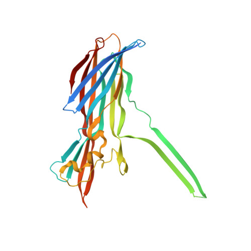
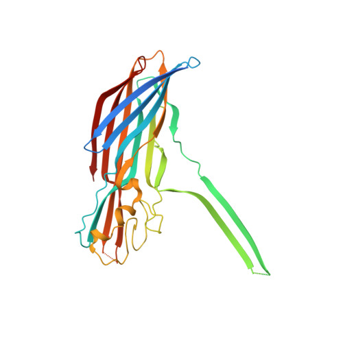
Molecular basis of transmembrane beta-barrel formation of staphylococcal pore-forming toxins.

Yamashita, D.Sugawara, T.Takeshita, M.Kaneko, J.Kamio, Y.Tanaka, I.Tanaka, Y.Yao, M.

(2014) Nat Commun 5: 4897-4897

  • DOI: https://doi.org/10.1038/ncomms5897
  • Primary Citation of Related Structures:  
    4P1X, 4P1Y

  • PubMed Abstract: 

    Pathogenic bacteria secrete pore-forming toxins (PFTs) to attack target cells. PFTs are expressed as water-soluble monomeric proteins, which oligomerize into nonlytic prepore intermediates on the target cell membrane before forming membrane-spanning pores. Despite a wealth of biochemical data, the lack of high-resolution prepore structural information has hampered understanding of the β-barrel formation process. Here, we report crystal structures of staphylococcal γ-haemolysin and leucocidin prepores. The structures reveal a disordered bottom half of the β-barrel corresponding to the transmembrane region, and a rigid upper extramembrane half. Spectroscopic analysis of fluorescently labelled mutants confirmed that the prepore is distinct from the pore within the transmembrane region. Mutational analysis also indicates a pivotal role for the glycine residue located at the lipid-solvent interface as a 'joint' between the two halves of the β-barrel. These observations suggest a two-step transmembrane β-barrel pore formation mechanism in which the upper extramembrane and bottom transmembrane regions are formed independently.


  • Organizational Affiliation

    Graduate School of Life Sciences, Hokkaido University, Sapporo 060-0810, Japan.


Macromolecules
Find similar proteins by:  (by identity cutoff)  |  3D Structure
Entity ID: 1
MoleculeChains Sequence LengthOrganismDetailsImage
Gamma-hemolysin component CA [auth B],
D,
F,
H
296Staphylococcus aureus subsp. aureus Mu50Mutation(s): 0 
Gene Names: hlgCSAV2420
Membrane Entity: Yes 
UniProt
Find proteins for Q99RL1 (Staphylococcus aureus (strain Mu50 / ATCC 700699))
Explore Q99RL1 
Go to UniProtKB:  Q99RL1
Entity Groups  
Sequence Clusters30% Identity50% Identity70% Identity90% Identity95% Identity100% Identity
UniProt GroupQ99RL1
Sequence Annotations
Expand
  • Reference Sequence
Find similar proteins by:  (by identity cutoff)  |  3D Structure
Entity ID: 2
MoleculeChains Sequence LengthOrganismDetailsImage
Gamma-hemolysin component BB [auth A],
C,
E,
G
309Staphylococcus aureus subsp. aureus Mu50Mutation(s): 1 
Gene Names: hlgBSAV2421
Membrane Entity: Yes 
UniProt
Find proteins for A0A0H3JX61 (Staphylococcus aureus (strain Mu50 / ATCC 700699))
Explore A0A0H3JX61 
Go to UniProtKB:  A0A0H3JX61
Entity Groups  
Sequence Clusters30% Identity50% Identity70% Identity90% Identity95% Identity100% Identity
UniProt GroupA0A0H3JX61
Sequence Annotations
Expand
  • Reference Sequence
Experimental Data & Validation

Experimental Data

  • Method: X-RAY DIFFRACTION
  • Resolution: 2.40 Å
  • R-Value Free: 0.223 
  • R-Value Work: 0.182 
  • R-Value Observed: 0.183 
  • Space Group: C 1 2 1
Unit Cell:
Length ( Å )Angle ( ˚ )
a = 193.31α = 90
b = 193.16β = 118.94
c = 116.38γ = 90
Software Package:
Software NamePurpose
PHENIXrefinement

Structure Validation

View Full Validation Report


Entry History 

Deposition Data

Revision History  (Full details and data files)

  • Version 1.0: 2014-10-01
    Type: Initial release
  • Version 1.1: 2014-10-15
    Changes: Database references
  • Version 2.0: 2023-12-27
    Changes: Advisory, Data collection, Database references, Derived calculations, Non-polymer description, Other, Refinement description, Source and taxonomy, Structure summary