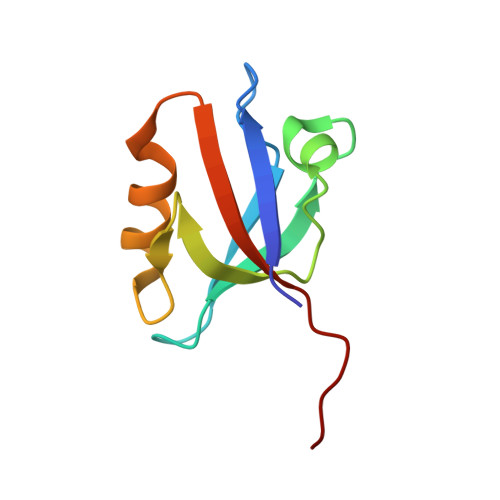
Structural insights into PDZ-mediated interaction of NHERF2 and LPA2, a cellular event implicated in CFTR channel regulation.
Holcomb, J., Jiang, Y., Lu, G., Trescott, L., Brunzelle, J., Sirinupong, N., Li, C., Naren, A.P., Yang, Z.(2014) Biochem Biophys Res Commun 446: 399-403
- PubMed: 24613836 Search on PubMedSearch on PubMed Central
- DOI: https://doi.org/10.1016/j.bbrc.2014.02.128
- Primary Citation Related Structures:
4P0C - PubMed Abstract:
The formation of CFTR-NHERF2-LPA2 macromolecular complex in airway epithelia regulates CFTR channel function and plays an important role in compartmentalized cAMP signaling. We previously have shown that disruption of the PDZ-mediated NHERF2-LPA2 interaction abolishes the LPA inhibitory effect and augments CFTR Cl(-) channel activity in vitro and in vivo. Here we report the first crystal structure of the NHERF2 PDZ1 domain in complex with the C-terminal LPA2 sequence. The structure reveals that the PDZ1-LPA2 binding specificity is achieved by numerous hydrogen bonds and hydrophobic contacts with the last four LPA2 residues contributing to specific interactions. Comparison of the PDZ1-LPA2 structure to the structure of PDZ1 in complex with a different peptide provides insights into the diverse nature of PDZ1 substrate recognition and suggests that the conformational flexibility in the ligand binding pocket is involved in determining the broad substrate specificity of PDZ1. In addition, the structure reveals a small surface pocket adjacent to the ligand-binding site, which may have therapeutic implications. This study provides an understanding of the structural basis for the PDZ-mediated NHERF2-LPA2 interaction that could prove valuable in selective drug design against CFTR-related human diseases.
- Department of Biochemistry and Molecular Biology, Wayne State University School of Medicine, Detroit, MI, USA.
Organizational Affiliation: