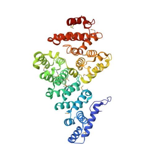
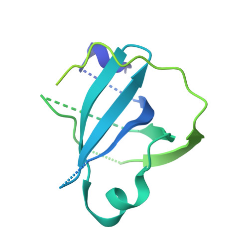
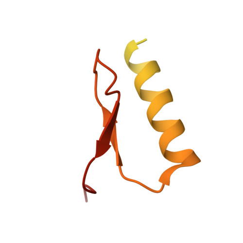
4OWT

Structural basis of SOSS1 complex assembly

  • Classification: DNA BINDING PROTEIN
  • Organism(s): Homo sapiens
  • Expression System: Escherichia coli
  • Mutation(s): No 

  • Deposited: 2014-02-03 Released: 2014-04-16 
  • Deposition Author(s): Ren, W., Sun, Q., Tang, X., Song, H.
  • Funding Organization(s): National Natural Science Foundation of China (NSFC), the Agency for Science, Technology and Research, National Basic Research Program of China (973 Program)

Experimental Data Snapshot

  • Method: X-RAY DIFFRACTION
  • Resolution: 2.00 Å
  • R-Value Free: 0.195 
  • R-Value Work: 0.172 
  • R-Value Observed: 0.173 

wwPDB Validation   3D Report Full Report


This is version 1.3 of the entry. See complete history


Literature

Structural Basis of SOSS1 Complex Assembly and Recognition of ssDNA.

Ren, W.Chen, H.Sun, Q.Tang, X.Lim, S.C.Huang, J.Song, H.

(2014) Cell Rep 6: 982-991

  • DOI: https://doi.org/10.1016/j.celrep.2014.02.020
  • Primary Citation of Related Structures:  
    4OWT, 4OWW, 4OWX

  • PubMed Abstract: 

    The SOSS1 complex comprising SOSSA, SOSSB1, and SOSSC senses single-stranded DNA (ssDNA) and promotes repair of DNA double-strand breaks (DSBs). But how SOSS1 is assembled and recognizes ssDNA remains elusive. The crystal structure of the N-terminal half of SOSSA (SOSSAN) in complex with SOSSB1 and SOSSC showed that SOSSAN serves as a scaffold to bind both SOSSB1 and SOSSC for assembly of the SOSS1 complex. The structures of SOSSAN/B1 in complex with a 12 nt ssDNA and SOSSAN/B1/C in complex with a 35 nt ssDNA showed that SOSSB1 interacts with both SOSSAN and ssDNA via two distinct surfaces. Recognition of ssDNA with a length of up to nine nucleotides is mediated solely by SOSSB1, whereas neither SOSSC nor SOSSAN are critical for ssDNA binding. These results reveal the structural basis of SOSS1 assembly and provide a framework for further study of the mechanism governing longer ssDNA recognition by the SOSS1 complex during DSB repair.


  • Organizational Affiliation

    Life Sciences Institute, Zhejiang University, 388 Yuhangtang Road, Hangzhou 310058, China; Institute of Molecular and Cell Biology, 61 Biopolis Drive, Singapore 138673, Singapore.


Macromolecules
Find similar proteins by:  (by identity cutoff)  |  3D Structure
Entity ID: 1
MoleculeChains Sequence LengthOrganismDetailsImage
Integrator complex subunit 3466Homo sapiensMutation(s): 0 
Gene Names: INTS3C1orf193C1orf60
UniProt & NIH Common Fund Data Resources
Find proteins for Q68E01 (Homo sapiens)
Explore Q68E01 
Go to UniProtKB:  Q68E01
GTEx:  ENSG00000143624 
Entity Groups  
Sequence Clusters30% Identity50% Identity70% Identity90% Identity95% Identity100% Identity
UniProt GroupQ68E01
Sequence Annotations
Expand
  • Reference Sequence
Find similar proteins by:  (by identity cutoff)  |  3D Structure
Entity ID: 2
MoleculeChains Sequence LengthOrganismDetailsImage
SOSS complex subunit B1211Homo sapiensMutation(s): 0 
Gene Names: NABP2OBFC2BSSB1LP3587
UniProt & NIH Common Fund Data Resources
Find proteins for Q9BQ15 (Homo sapiens)
Explore Q9BQ15 
Go to UniProtKB:  Q9BQ15
PHAROS:  Q9BQ15
GTEx:  ENSG00000139579 
Entity Groups  
Sequence Clusters30% Identity50% Identity70% Identity90% Identity95% Identity100% Identity
UniProt GroupQ9BQ15
Sequence Annotations
Expand
  • Reference Sequence
Find similar proteins by:  (by identity cutoff)  |  3D Structure
Entity ID: 3
MoleculeChains Sequence LengthOrganismDetailsImage
SOSS complex subunit C104Homo sapiensMutation(s): 0 
Gene Names: INIPC9orf80SSBIP1HSPC043HSPC291
UniProt & NIH Common Fund Data Resources
Find proteins for Q9NRY2 (Homo sapiens)
Explore Q9NRY2 
Go to UniProtKB:  Q9NRY2
PHAROS:  Q9NRY2
GTEx:  ENSG00000148153 
Entity Groups  
Sequence Clusters30% Identity50% Identity70% Identity90% Identity95% Identity100% Identity
UniProt GroupQ9NRY2
Sequence Annotations
Expand
  • Reference Sequence
Small Molecules
Modified Residues  1 Unique
IDChains TypeFormula2D DiagramParent
MSE
Query on MSE
A
L-PEPTIDE LINKINGC5 H11 N O2 SeMET
Experimental Data & Validation

Experimental Data

  • Method: X-RAY DIFFRACTION
  • Resolution: 2.00 Å
  • R-Value Free: 0.195 
  • R-Value Work: 0.172 
  • R-Value Observed: 0.173 
  • Space Group: P 31 2 1
Unit Cell:
Length ( Å )Angle ( ˚ )
a = 116.809α = 90
b = 116.809β = 90
c = 109.832γ = 120
Software Package:
Software NamePurpose
PHENIXrefinement

Structure Validation

View Full Validation Report


Entry History & Funding Information

Deposition Data


Funding OrganizationLocationGrant Number
National Natural Science Foundation of China (NSFC)China31270816
the Agency for Science, Technology and ResearchSingapore--
National Basic Research Program of China (973 Program)China2012CB944402
National Basic Research Program of China (973 Program)China2013CB911003

Revision History  (Full details and data files)

  • Version 1.0: 2014-04-16
    Type: Initial release
  • Version 1.1: 2017-09-06
    Changes: Advisory, Author supporting evidence, Derived calculations, Other, Source and taxonomy, Structure summary
  • Version 1.2: 2020-01-08
    Changes: Author supporting evidence
  • Version 1.3: 2023-12-27
    Changes: Data collection, Database references, Refinement description