4OQ9 | pdb_00004oq9

Satellite Tobacco Mosaic Virus Refined to 1.4 A Resolution using non-crystallographic symmetry restraints


Experimental Data Snapshot

  • Method: X-RAY DIFFRACTION
  • Resolution: 1.45 Å
  • R-Value Free: 
    0.250 (Depositor), 0.282 (DCC) 
  • R-Value Work: 
    0.217 (Depositor) 
  • R-Value Observed: 
    0.218 (Depositor) 

Starting Model: experimental
View more details

wwPDB Validation 3D Report Full Report

Validation slider image for 4OQ9

This is version 1.2 of the entry. See complete history

Literature

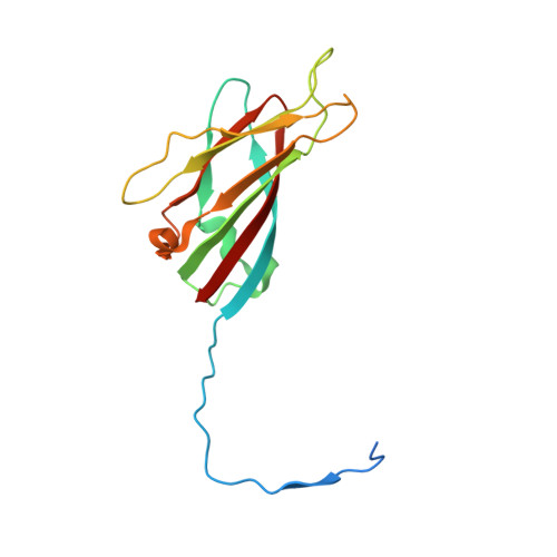
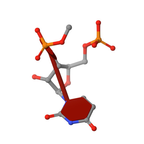
Satellite tobacco mosaic virus refined to 1.4 angstrom resolution.

Larson, S.B.Day, J.S.McPherson, A.

(2014) Acta Crystallogr D Biol Crystallogr 70: 2316-2330

  • DOI: https://doi.org/10.1107/S1399004714013789
  • Primary Citation Related Structures: 
    4NIA, 4OQ8, 4OQ9

  • PubMed Abstract: 

    Satellite tobacco mosaic virus (STMV) is among the smallest viruses, having 60 identical subunits arranged with T = 1 icosahedral symmetry. Its crystal structure was solved at 290 K and was refined using, in part, crystals grown in microgravity. Electron-density maps revealed nearly 57% of the genomic ssRNA. Using six flash-cooled crystals, diffraction data were recorded to 1.4 Å resolution and independent refinements of the STMV model were carried out versus the previous 1.8 Å resolution data representing merged data from 21 crystals (271,689 unique reflections), data consisting of corresponding reflections to 1.8 Å resolution from the cooled crystals and 1.4 Å resolution data from the cooled crystals comprised of 570,721 unique reflections. Models were independently refined with full NCS constraints using the program CNS and in restrained mode using the programs CNS, REFMAC5 and SHELX-97, with the latter two procedures including anisotropic temperature factors. Significant additional structural detail emerged from the analyses, including a unique cation and anion arrangement on fivefold axes and a precise assessment of icosahedral symmetry exactness in the crystal lattice. STMV represents the highest resolution native virus structure currently known by a substantial margin, and it permits the evaluation of a precise atomic model of a spherical virus at near-atomic resolution for the first time.


  • Organizational Affiliation
    • Department of Molecular Biology and Biochemistry, University of California, Irvine, Irvine, CA 92697-3900, USA.

Macromolecule Content 

  • Total Structure Weight: 367.95 kDa 
  • Atom Count: 29,296 
  • Modeled Residue Count: 2,490 
  • Deposited Residue Count: 2,715 
  • Unique protein chains: 1
  • Unique nucleic acid chains: 3

Macromolecules


Find similar proteins by:|  3D Structure
Entity ID: 1
MoleculeChains  Sequence LengthOrganismDetailsImage
Coat protein
A, B, C, D, E
A, B, C, D, E, F, G, H, I, J, K, L, M, N, O
159Satellite tobacco mosaic virusMutation(s): 0 
UniProt
Find proteins for P17574 (Satellite tobacco mosaic virus)
Explore P17574 
Go to UniProtKB:  P17574
Entity Groups
Sequence Clusters30% Identity50% Identity70% Identity90% Identity95% Identity100% Identity
UniProt GroupP17574
Sequence Annotations
Expand
Reference Sequence
Find similar nucleic acids by:  Sequence
Entity ID: 2
MoleculeChains LengthOrganismImage
RNA (5'-R(P*AP*AP*AP*AP*AP*AP*AP*AP*AP*A)-3')10Satellite tobacco mosaic virus
Sequence Annotations
Expand
Reference Sequence
Find similar nucleic acids by:  Sequence
Entity ID: 3
MoleculeChains LengthOrganismImage
RNA (5'-R(P*UP*UP*UP*UP*UP*UP*UP*UP*UP*U)-3')10Satellite tobacco mosaic virus
Sequence Annotations
Expand
Reference Sequence
Find similar nucleic acids by:  Sequence
Entity ID: 4
MoleculeChains LengthOrganismImage
RNA (5'-R(P*UP*U)-3')2Satellite tobacco mosaic virus
Sequence Annotations
Expand
Reference Sequence

Small Molecules

Ligands 4 Unique
IDChains Name / Formula / InChI Key2D Diagram3D Interactions
SO4

Query on SO4


Download:Ideal Coordinates CCD File
AC [auth G]
BC [auth G]
EC [auth H]
GC [auth I]
IC [auth J]
AC [auth G],
BC [auth G],
EC [auth H],
GC [auth I],
IC [auth J],
JB [auth A],
KC [auth K],
LB [auth B],
NC [auth L],
OB [auth C],
PC [auth M],
QB [auth D],
QC [auth M],
SB [auth E],
TB [auth E],
TC [auth N],
VC [auth O],
XB [auth F]
SULFATE ION
O4 S
QAOWNCQODCNURD-UHFFFAOYSA-L
PO4

Query on PO4


Download:Ideal Coordinates CCD File
DC [auth H],
IB [auth A],
SC [auth N]
PHOSPHATE ION
O4 P
NBIIXXVUZAFLBC-UHFFFAOYSA-K
MG

Query on MG


Download:Ideal Coordinates CCD File
LC [auth K],
UB [auth E],
YB [auth F]
MAGNESIUM ION
Mg
JLVVSXFLKOJNIY-UHFFFAOYSA-N
NA

Query on NA


Download:Ideal Coordinates CCD File
CC [auth G]
FC [auth H]
HC [auth I]
JC [auth J]
KB [auth A]
CC [auth G],
FC [auth H],
HC [auth I],
JC [auth J],
KB [auth A],
MB [auth B],
MC [auth K],
NB [auth B],
OC [auth L],
PB [auth C],
RB [auth D],
RC [auth M],
UC [auth N],
VB [auth E],
WB [auth E],
WC [auth O],
ZB [auth F]
SODIUM ION
Na
FKNQFGJONOIPTF-UHFFFAOYSA-N

Experimental Data & Validation

Experimental Data

  • Method: X-RAY DIFFRACTION
  • Resolution: 1.45 Å
  • R-Value Free:  0.250 (Depositor), 0.282 (DCC) 
  • R-Value Work:  0.217 (Depositor) 
  • R-Value Observed: 0.218 (Depositor) 
Space Group: I 2 2 2
Unit Cell:
Length ( Å )Angle ( ˚ )
a = 172.69α = 90
b = 190.3β = 90
c = 201.69γ = 90
Software Package:
Software NamePurpose
ADSCdata collection
Cootmodel building
REFMACrefinement
d*TREKdata reduction
d*TREKdata scaling
CNSphasing

Structure Validation

View Full Validation Report


Entry History 

Deposition Data

Revision History  (Full details and data files)

  • Version 1.0: 2014-09-10
    Type: Initial release
  • Version 1.1: 2014-10-01
    Changes: Database references
  • Version 1.2: 2023-09-20
    Changes: Data collection, Database references, Derived calculations, Refinement description