4OQ3 | pdb_00004oq3

Tetra-substituted imidazoles as a new class of inhibitors of the p53-MDM2 interaction


Experimental Data Snapshot

  • Method: X-RAY DIFFRACTION
  • Resolution: 2.30 Å
  • R-Value Free: 
    0.224 (Depositor), 0.223 (DCC) 
  • R-Value Work: 
    0.194 (Depositor), 0.195 (DCC) 
  • R-Value Observed: 
    0.196 (Depositor) 

Starting Model: experimental
View more details

wwPDB Validation 3D Report Full Report

Validation slider image for 4OQ3

Ligand Structure Quality Assessment 


This is version 1.3 of the entry. See complete history

Literature

Tetra-substituted imidazoles as a new class of inhibitors of the p53-MDM2 interaction.

Vaupel, A.Bold, G.De Pover, A.Stachyra-Valat, T.Hergovich-Lisztwan, J.Kallen, J.Masuya, K.Furet, P.

(2014) Bioorg Med Chem Lett 24: 2110-2114

  • DOI: https://doi.org/10.1016/j.bmcl.2014.03.039
  • Primary Citation Related Structures: 
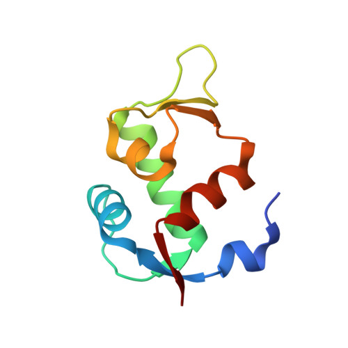
    4OQ3

  • PubMed Abstract: 

    Capitalizing on crystal structure information obtained from a previous effort in the search for non peptide inhibitors of the p53-MDM2 interaction, we have discovered another new class of compounds able to disrupt this protein-protein interaction, an important target in oncology drug research. The new inhibitors, based on a tetra-substituted imidazole scaffold, have been optimized to low nanomolar potency in a biochemical assay following a structure-guided approach. An appropriate strategy has allowed us to translate the high biochemical potency in significant anti-proliferative activity on a p53-dependent MDM2 amplified cell line.


  • Organizational Affiliation
    • Novartis Institutes for BioMedical Research, CH-4002 Basel, Switzerland. Electronic address: andrea.vaupel@novartis.com.

Macromolecule Content 

  • Total Structure Weight: 46.44 kDa 
  • Atom Count: 3,343 
  • Modeled Residue Count: 367 
  • Deposited Residue Count: 384 
  • Unique protein chains: 1

Macromolecules

Find similar proteins by:|  3D Structure
Entity ID: 1
MoleculeChains  Sequence LengthOrganismDetailsImage
E3 ubiquitin-protein ligase Mdm2
A, B, C, D
96Homo sapiensMutation(s): 1 
Gene Names: MDM2
EC: 6.3.2 (PDB Primary Data), 2.3.2.27 (UniProt)
UniProt & NIH Common Fund Data Resources
Find proteins for Q00987 (Homo sapiens)
Explore Q00987 
Go to UniProtKB:  Q00987
PHAROS:  Q00987
GTEx:  ENSG00000135679 
Entity Groups
Sequence Clusters30% Identity50% Identity70% Identity90% Identity95% Identity100% Identity
UniProt GroupQ00987
Sequence Annotations
Expand
Reference Sequence

Experimental Data & Validation

Experimental Data

  • Method: X-RAY DIFFRACTION
  • Resolution: 2.30 Å
  • R-Value Free:  0.224 (Depositor), 0.223 (DCC) 
  • R-Value Work:  0.194 (Depositor), 0.195 (DCC) 
  • R-Value Observed: 0.196 (Depositor) 
Space Group: P 1 21 1
Unit Cell:
Length ( Å )Angle ( ˚ )
a = 33.814α = 90
b = 165.528β = 95.06
c = 34.236γ = 90
Software Package:
Software NamePurpose
DENZOdata reduction
SCALEPACKdata scaling
PHASERphasing
REFMACrefinement
PDB_EXTRACTdata extraction

Structure Validation

View Full Validation Report


Ligand Structure Quality Assessment 


Entry History 

Deposition Data

  • Released Date: 2014-04-23 
  • Deposition Author(s): Kallen, J.

Revision History  (Full details and data files)

  • Version 1.0: 2014-04-23
    Type: Initial release
  • Version 1.1: 2014-05-21
    Changes: Database references
  • Version 1.2: 2017-11-22
    Changes: Refinement description
  • Version 1.3: 2023-09-20
    Changes: Data collection, Database references, Derived calculations, Refinement description