4OQ1 | pdb_00004oq1

Structure of the Streptococcal ancillary pilin


Experimental Data Snapshot

  • Method: X-RAY DIFFRACTION
  • Resolution: 1.85 Å
  • R-Value Free: 
    0.237 (Depositor), 0.246 (DCC) 
  • R-Value Work: 
    0.186 (Depositor), 0.189 (DCC) 
  • R-Value Observed: 
    0.188 (Depositor) 

wwPDB Validation 3D Report Full Report

Validation slider image for 4OQ1

This is version 1.3 of the entry. See complete history

Literature

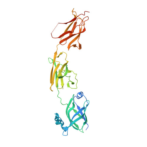
Structural Basis of Pilus Anchoring by the Ancillary Pilin RrgC of Streptococcus pneumoniae.

Shaik, M.M.Maccagni, A.Tourcier, G.Di Guilmi, A.M.Dessen, A.

(2014) J Biological Chem 289: 16988-16997

  • DOI: https://doi.org/10.1074/jbc.M114.555854
  • Primary Citation Related Structures: 
    4OQ1

  • PubMed Abstract: 

    Pili are surface-attached, fibrous virulence factors that play key roles in the pathogenesis process of a number of bacterial agents. Streptococcus pneumoniae is a causative agent of pneumonia and meningitis, and the appearance of drug-resistance organisms has made its treatment challenging, especially in developing countries. Pneumococcus-expressed pili are composed of three structural proteins: RrgB, which forms the polymerized backbone, RrgA, the tip-associated adhesin, and RrgC, which presumably associates the pilus with the bacterial cell wall. Despite the fact that the structures of both RrgA and RrgB were known previously, structural information for RrgC was still lacking, impeding the analysis of a complete model of pilus architecture. Here, we report the structure of RrgC to 1.85 Å and reveal that it is a three-domain molecule stabilized by two intradomain isopeptide bonds. RrgC does not depend on pilus-specific sortases to become attached to the cell wall; instead, it binds the preformed pilus to the peptidoglycan by employing the catalytic activity of SrtA. A comprehensive model of the type 1 pilus from S. pneumoniae is also presented.


  • Organizational Affiliation
    • From the Institut de Biologie Structurale (IBS), Université Grenoble Alpes, 41 avenue des Martyrs, 38044 Grenoble, France, the Commissariat à l'Energie Atomique (CEA), 38000 Grenoble, France, the Centre National de la Recherche Scientifique (CNRS), UMR 5075, Grenoble, France, and.

Macromolecule Content 

  • Total Structure Weight: 41.97 kDa 
  • Atom Count: 2,884 
  • Modeled Residue Count: 331 
  • Deposited Residue Count: 368 
  • Unique protein chains: 1

Macromolecules

Find similar proteins by:|  3D Structure
Entity ID: 1
MoleculeChains  Sequence LengthOrganismDetailsImage
Cell wall surface anchor family protein368Streptococcus pneumoniae TIGR4Mutation(s): 3 
Gene Names: SP0464SP_0464
UniProt
Find proteins for A0A0H2UNM0 (Streptococcus pneumoniae serotype 4 (strain ATCC BAA-334 / TIGR4))
Explore A0A0H2UNM0 
Go to UniProtKB:  A0A0H2UNM0
Entity Groups
Sequence Clusters30% Identity50% Identity70% Identity90% Identity95% Identity100% Identity
UniProt GroupA0A0H2UNM0
Sequence Annotations
Expand
Reference Sequence

Experimental Data & Validation

Experimental Data

  • Method: X-RAY DIFFRACTION
  • Resolution: 1.85 Å
  • R-Value Free:  0.237 (Depositor), 0.246 (DCC) 
  • R-Value Work:  0.186 (Depositor), 0.189 (DCC) 
  • R-Value Observed: 0.188 (Depositor) 
Space Group: P 1 21 1
Unit Cell:
Length ( Å )Angle ( ˚ )
a = 43.58α = 90
b = 65.76β = 110.97
c = 60.42γ = 90
Software Package:
Software NamePurpose
MxCuBEdata collection
SHARPphasing
PHENIXrefinement
MOSFLMdata reduction
SCALAdata scaling

Structure Validation

View Full Validation Report


Entry History 

Deposition Data

Revision History  (Full details and data files)

  • Version 1.0: 2014-04-23
    Type: Initial release
  • Version 1.1: 2014-06-04
    Changes: Database references
  • Version 1.2: 2014-07-02
    Changes: Database references
  • Version 1.3: 2024-11-06
    Changes: Data collection, Database references, Derived calculations, Structure summary